Deposition Date
2000-11-27
Release Date
2002-08-28
Last Version Date
2024-04-03
Entry Detail
PDB ID:
1G9S
Keywords:
Title:
CRYSTAL STRUCTURE OF A COMPLEX BETWEEN E.COLI HPRT AND IMP
Biological Source:
Source Organism(s):
Escherichia coli (Taxon ID: 562)
Expression System(s):
Method Details:
Experimental Method:
Resolution:
2.80 Å
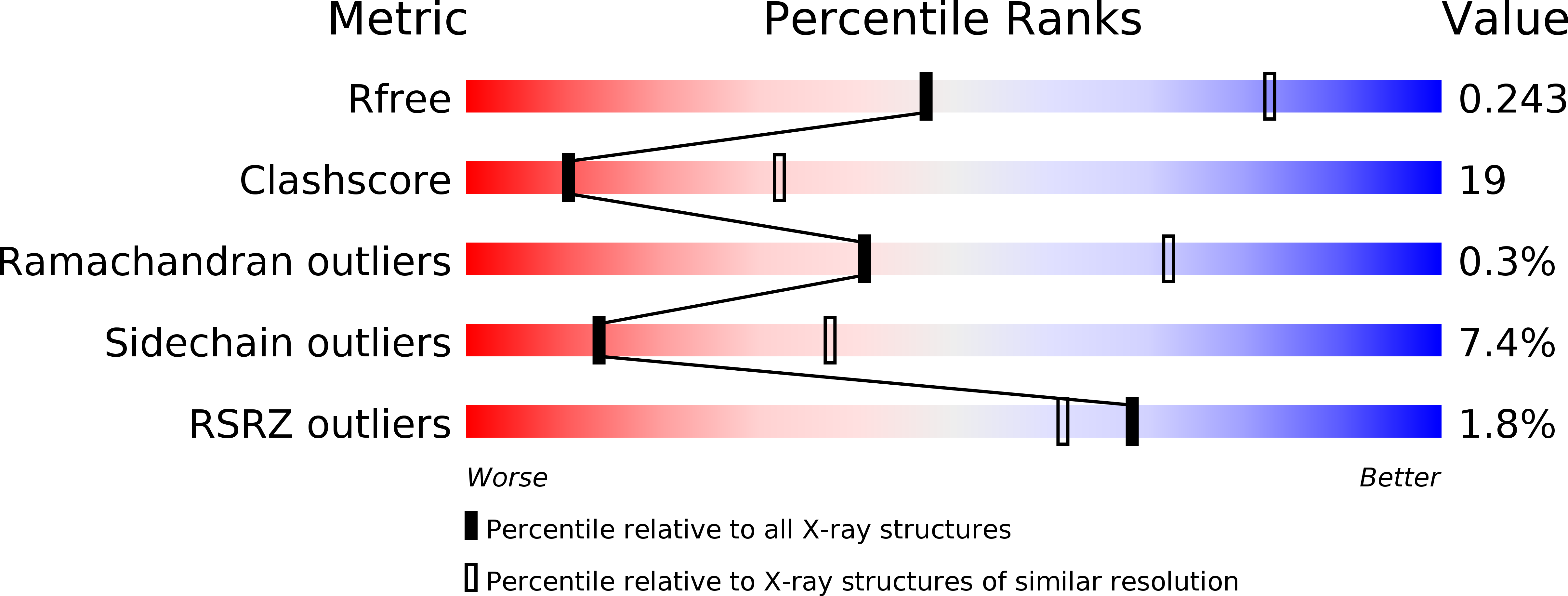
R-Value Free:
0.24
R-Value Work:
0.20
R-Value Observed:
0.20
Space Group:
P 31 2 1