1G97 image
Deposition Date 2000-11-22
Release Date 2001-05-22
Last Version Date 2023-08-09
Entry Detail
PDB ID:
1G97
Keywords:
Title:
S.PNEUMONIAE GLMU COMPLEXED WITH UDP-N-ACETYLGLUCOSAMINE AND MG2+
Biological Source:
Source Organism:
Host Organism:
Method Details:
Experimental Method:
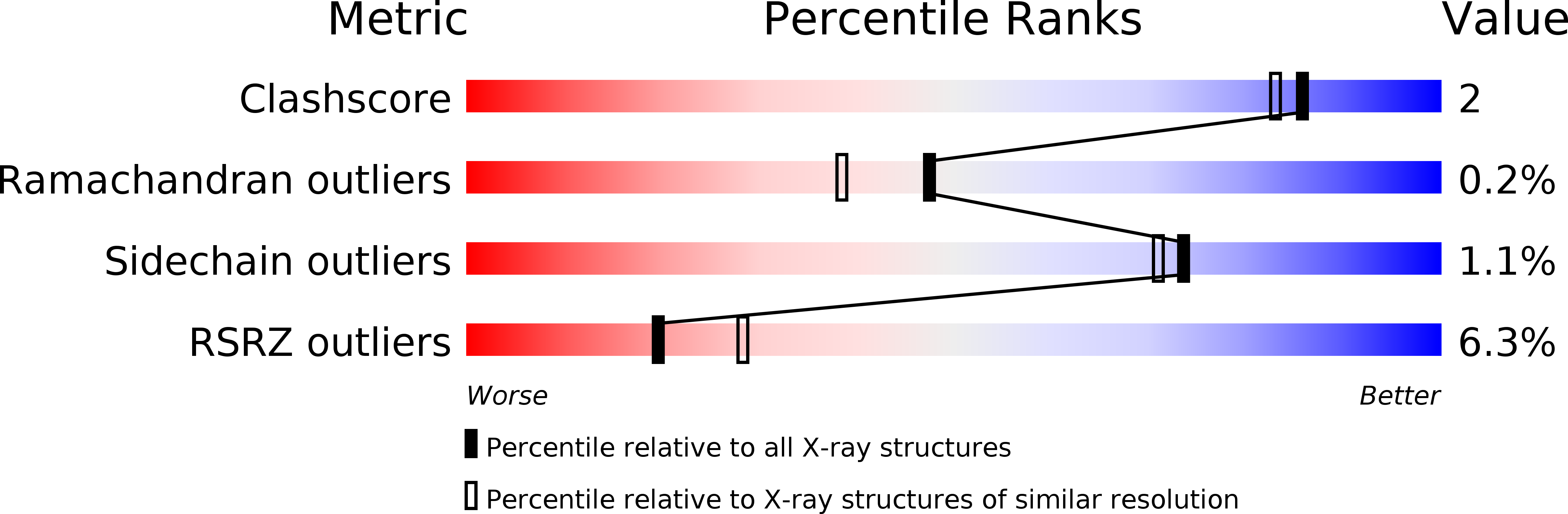
Resolution:
1.96 Å
R-Value Free:
0.22
R-Value Work:
0.18
Space Group:
H 3 2
Macromolecular Entities
Structures with similar UniProt ID
Protein Blast
Polymer Type:polypeptide(L)
Molecule:N-ACETYLGLUCOSAMINE-1-PHOSPHATE URIDYLTRANSFERASE
Gene (Uniprot):glmU
Chain IDs:A
Chain Length:459
Number of Molecules:1
Biological Source:Streptococcus pneumoniae
Primary Citation
Crystal structures of Streptococcus pneumoniae N-acetylglucosamine-1-phosphate uridyltransferase, GlmU, in apo form at 2.33 A resolution and in complex with UDP-N-acetylglucosamine and Mg(2+) at 1.96 A resolution.
J.Mol.Biol. 305 279 289 (2001)
PMID: 11124906 DOI: 10.1006/jmbi.2000.4296

Abstact

N-Acetylglucosamine-1-phosphate uridyltransferase (GlmU) is an essential bacterial enzyme with both an acetyltransferase and a uridyltransferase activity which have been mapped to the C-terminal and N-terminal domains, respectively. GlmU performs the last two steps in the synthesis of UDP-N-acetylglucosamine (UDP-GlcNAc), which is an essential precursor in both the peptidoglycan and the lipopolysaccharide metabolic pathways. GlmU is therefore an attractive target for potential antibiotics. Knowledge of its three-dimensional structure would provide a basis for rational drug design. We have determined the crystal structures of Streptococcus pneumoniae GlmU (SpGlmU) in apo form at 2.33 A resolution, and in complex with UDP-N-acetyl glucosamine and the essential co-factor Mg(2+) at 1.96 A resolution. The protein structure consists of an N-terminal domain with an alpha/beta-fold, containing the uridyltransferase active site, and a C-terminal domain with a long left-handed beta-sheet helix (LbetaH) domain. An insertion loop containing the highly conserved sequence motif Asn-Tyr-Asp-Gly protrudes from the left-handed beta-sheet helix domain. In the crystal, S. pneumoniae GlmU forms exact trimers, mainly through contacts between left-handed beta-sheet helix domains. UDP-N-acetylglucosamine and Mg(2+) are bound at the uridyltransferase active site, which is in a closed form. We propose a uridyltransferase mechanism in which the activation energy of the double negatively charged phosphorane transition state is lowered by charge compensation of Mg(2+) and the side-chain of Lys22.

Legend

Protein

Chemical

Disease

Primary Citation of related structures
Feedback Form
Name
Email
Institute
Feedback