
Deposition Date
2000-11-16
Release Date
2000-11-29
Last Version Date
2024-11-13
Entry Detail
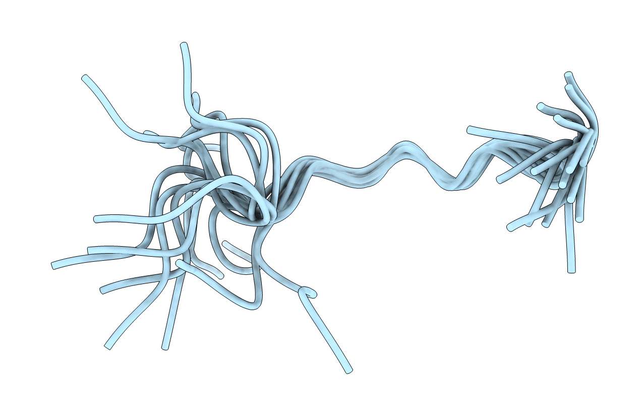
PDB ID:
1G8C
Keywords:
Title:
STRUCTURE OF THE BOVINE ANTIMICROBIAL PEPTIDE INDOLICIDIN BOUND TO SODIUM DODECYL SULFATE MICELLES
Method Details:
Experimental Method:
Conformers Calculated:
100
Conformers Submitted:
16
Selection Criteria:
structures with the least restraint violations,structures with the lowest energy
