
Deposition Date
2000-11-14
Release Date
2001-05-16
Last Version Date
2024-02-07
Entry Detail
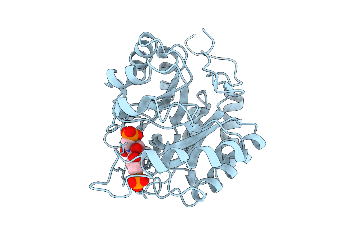
PDB ID:
1G7V
Keywords:
Title:
CRYSTAL STRUCTURES OF KDO8P SYNTHASE IN ITS BINARY COMPLEXES WITH THE MECHANISM-BASED INHIBITOR
Biological Source:
Source Organism(s):
Escherichia coli (Taxon ID: 562)
Expression System(s):
Method Details:
Experimental Method:
Resolution:
2.40 Å
R-Value Free:
0.24
R-Value Work:
0.22
R-Value Observed:
0.22
Space Group:
I 2 3
