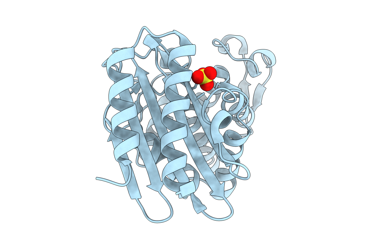
1G68 image
Entry Detail
PDB ID:
1G68
Keywords:
Title:
PSE-4 CARBENICILLINASE, WILD TYPE
Biological Source:
Source Organism:
Host Organism:
PDB Version:
Deposition Date:
2000-11-03
Release Date:
2001-02-21
Method Details:
Experimental Method:
Resolution:
1.95 Å
R-Value Free:
0.21
R-Value Work:
0.16
R-Value Observed:
0.16
Space Group:
P 41 21 2
Macromolecular Entities
Polymer Type:polypeptide(L)
Description:BETA-LACTAMASE PSE-4
Chain IDs:A
Chain Length:271
Number of Molecules:1
Biological Source:Pseudomonas aeruginosa
Ligand Molecules
Primary Citation
Insights into the molecular basis for the carbenicillinase activity of PSE-4 beta-lactamase from crystallographic and kinetic studies.
Biochemistry 40 395 402 (2001)
PMID: 11148033 DOI: 10.1021/bi001653v

Abstact

PSE-4 is a class A beta-lactamase produced by strains of Pseudomonas aeruginosa and is highly active for the penicillin derivative carbenicillin. The crystal structure of the wild-type PSE-4 carbenicillinase has been determined to 1.95 A resolution by molecular replacement and represents the first structure of a carbenicillinase published to date. A superposition of the PSE-4 structure with that of TEM-1 shows a rms deviation of 1.3 A for 263 Calpha atoms. Most carbenicillinases are unique among class A beta-lactamases in that residue 234 is an arginine (ABL standard numbering scheme), while in all other class A enzymes this residue is a lysine. Kinetic characterization of a R234K PSE-4 mutant reveals a 50-fold reduction in k(cat)/K(m) and confirms the importance of Arg 234 for carbenicillinase activity. A comparison of the structure of the R234K mutant refined to 1.75 A resolution with the wild-type structure shows that Arg 234 stabilizes an alternate conformation of the Ser 130 side chain, not seen in other class A beta-lactamase structures. Our molecular modeling studies suggest that the position of a bound carbenicillin would be shifted relative to that of a bound benzylpenicillin in order to avoid a steric clash between the carbenicillin alpha-carboxylate group and the conserved side chain of Asn 170. The alternate conformation of the catalytic Ser 130 in wild-type PSE-4 may be involved in accommodating this shift in the bound substrate position.

Legend

Protein

Chemical

Disease

Primary Citation of related structures