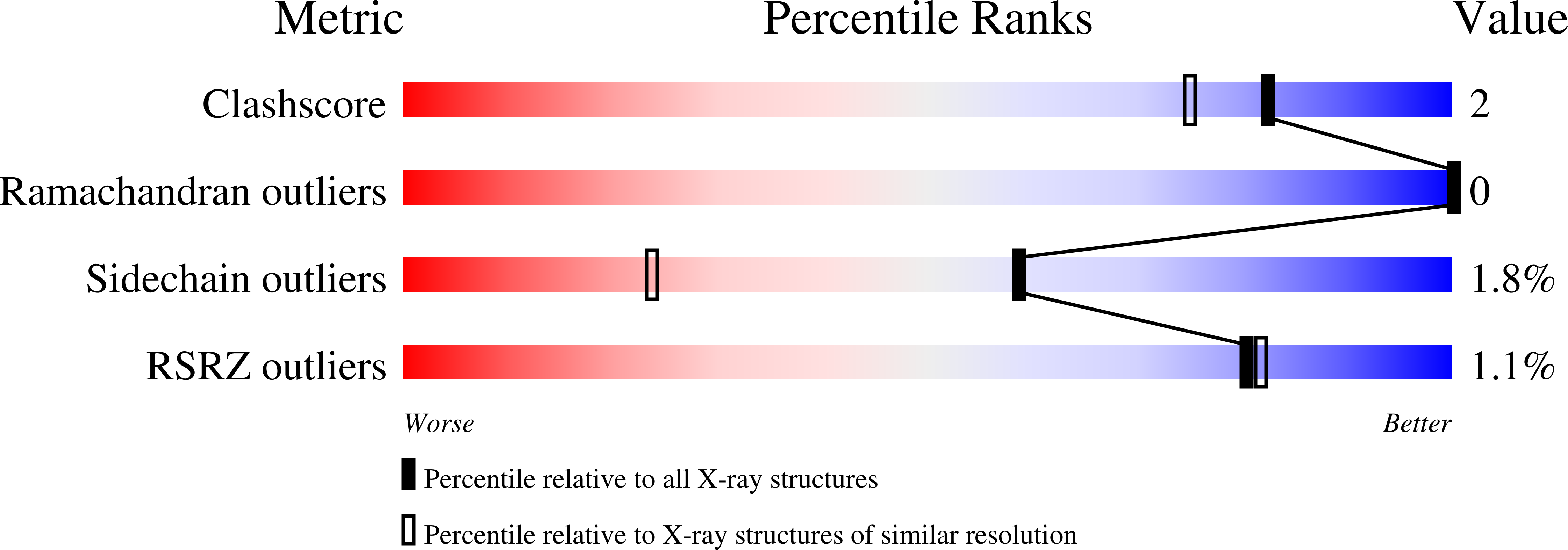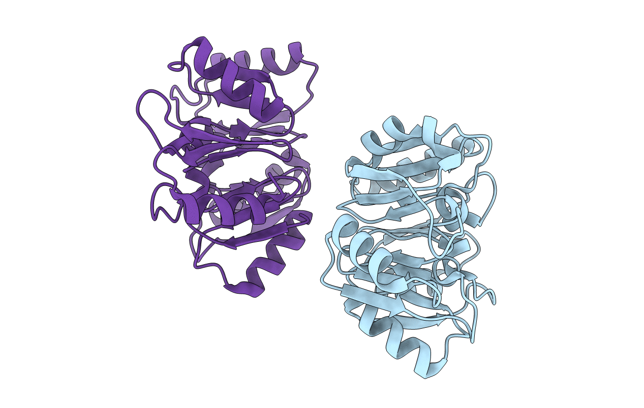
1G61 image
Deposition Date 2000-11-02
Release Date 2000-11-22
Last Version Date 2024-02-07
Entry Detail
PDB ID:
1G61
Keywords:
Title:
CRYSTAL STRUCTURE OF M.JANNASCHII EIF6
Biological Source:
Source Organism:
Host Organism:
Method Details:
Experimental Method:
Resolution:
1.30 Å
R-Value Free:
0.17
R-Value Work:
0.13
R-Value Observed:
0.13
Space Group:
C 1 2 1
Macromolecular Entities
Structures with similar UniProt ID
Protein Blast
Polymer Type:polypeptide(L)
Molecule:TRANSLATION INITIATION FACTOR 6
Gene (Uniprot):eif6
Chain IDs:A, B
Chain Length:228
Number of Molecules:2
Biological Source:Methanocaldococcus jannaschii
Primary Citation
Crystal structures of ribosome anti-association factor IF6.
Nat.Struct.Biol. 7 1156 1164 (2000)
PMID: 11101899 DOI: 10.1038/82017

Abstact

Ribosome anti-association factor eIF6 (originally named according to translation initiation terminology as eukaryotic initiation factor 6) binds to the large ribosomal subunit, thereby preventing inappropriate interactions with the small subunit during initiation of protein synthesis. We have determined the X-ray structures of two IF6 homologs, Methanococcus jannaschii archaeal aIF6 and Sacchromyces cerevisiae eIF6, revealing a phylogenetically conserved 25 kDa protein consisting of five quasi identical alpha/beta subdomains arrayed about a five-fold axis of pseudosymmetry. Yeast eIF6 prevents ribosomal subunit association. Comparative protein structure modeling with other known archaeal and eukaryotic homologs demonstrated the presence of two conserved surface regions, one or both of which may bind the large ribosomal subunit.

Legend

Protein

Chemical

Disease

Primary Citation of related structures