Deposition Date
2000-11-02
Release Date
2002-05-01
Last Version Date
2024-02-07
Entry Detail
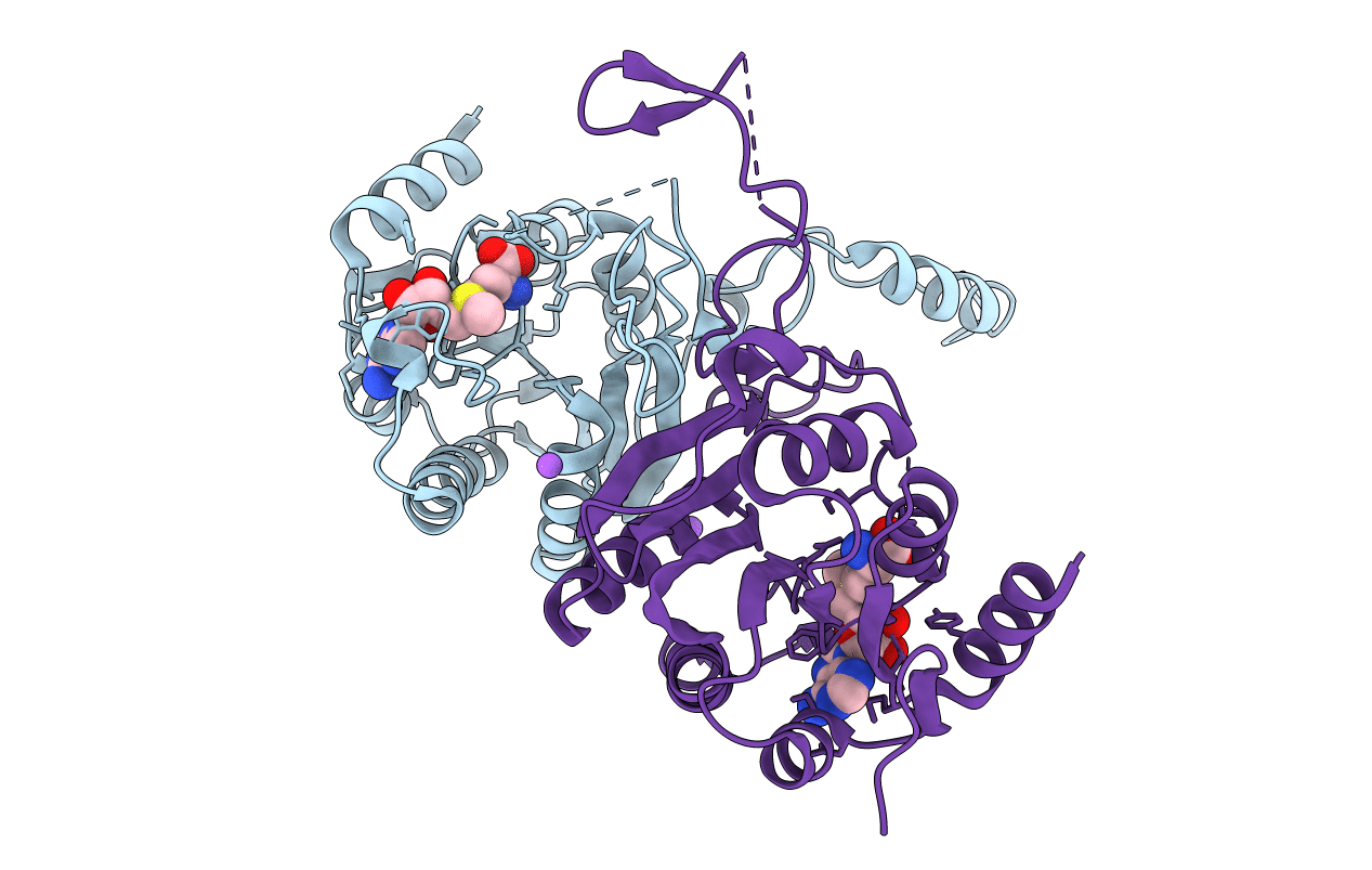
PDB ID:
1G60
Keywords:
Title:
Crystal Structure of Methyltransferase MboIIa (Moraxella bovis)
Biological Source:
Source Organism(s):
Moraxella bovis (Taxon ID: 476)
Expression System(s):
Method Details:
Experimental Method:
Resolution:
1.74 Å
R-Value Free:
0.22
R-Value Work:
0.19
R-Value Observed:
0.19
Space Group:
P 1 21 1