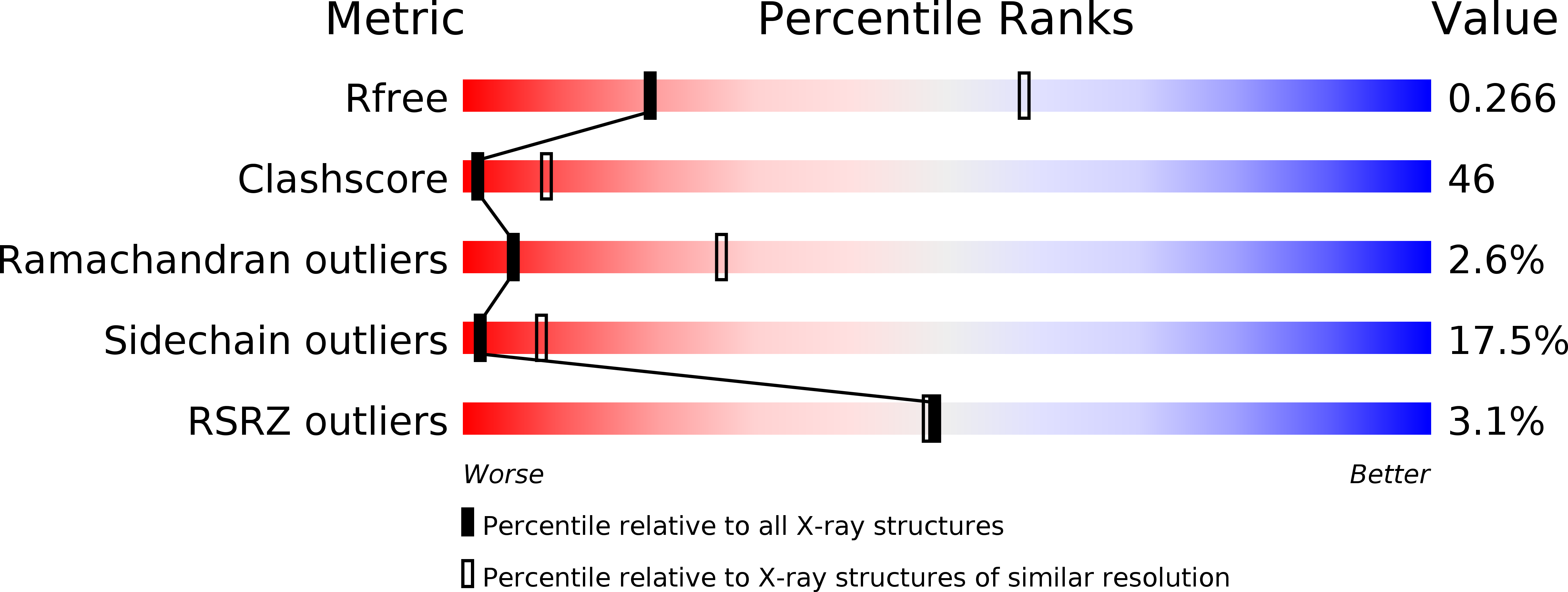
1G5G image
Deposition Date 2000-11-01
Release Date 2002-02-27
Last Version Date 2024-11-20
Entry Detail
PDB ID:
1G5G
Keywords:
Title:
FRAGMENT OF FUSION PROTEIN FROM NEWCASTLE DISEASE VIRUS
Biological Source:
Source Organism:
Method Details:
Experimental Method:
Resolution:
3.30 Å
R-Value Free:
0.27
R-Value Work:
0.22
Space Group:
C 2 2 21
Macromolecular Entities
Structures with similar UniProt ID
Protein Blast
Polymer Type:polypeptide(L)
Molecule:Fusion glycoprotein F0
Chain IDs:A, B, C, D, E, F
Chain Length:481
Number of Molecules:6
Biological Source:Avian orthoavulavirus 1
Modified Residue
Compound ID Chain ID Parent Comp ID Details 2D Image
ASN A ASN GLYCOSYLATION SITE
Ligand Molecules
Primary Citation
The structure of the fusion glycoprotein of Newcastle disease virus suggests a novel paradigm for the molecular mechanism of membrane fusion.
Structure 9 255 266 (2001)
PMID: 11286892 DOI: 10.1016/S0969-2126(01)00581-0

Abstact

BACKGROUND: Membrane fusion within the Paramyxoviridae family of viruses is mediated by a surface glycoprotein termed the "F", or fusion, protein. Membrane fusion is assumed to involve a series of structural transitions of F from a metastable (prefusion) state to a highly stable (postfusion) state. No detail is available at the atomic level regarding the metastable form of these proteins or regarding the transitions accompanying fusion. RESULTS: The three-dimensional structure of the fusion protein of Newcastle disease virus (NDV-F) has been determined. The trimeric NDV-F molecule is organized into head, neck, and stalk regions. The head is comprised of a highly twisted beta domain and an additional immunoglobulin-like beta domain. The neck is formed by the C-terminal extension of the heptad repeat region HR-A, capped by a four-helical bundle. The C terminus of HR-A is encased by a further helix HR-C and a 4-stranded beta sheet. The stalk is formed by the remaining visible portion of HR-A and by polypeptide immediately N-terminal to the C-terminal heptad repeat region HR-B. An axial channel extends through the head and neck and is fenestrated by three large radial channels located approximately at the head-neck interface. CONCLUSION: We propose that prior to fusion activation, the hydrophobic fusion peptides in NDV-F are sequestered within the radial channels within the head, with the central HR-A coiled coil being only partly formed. Fusion activation then involves, inter alia, the assembly of a complete HR-A coiled coil, with the fusion peptides and transmembrane anchors being brought into close proximity. The structure of NDV-F is fundamentally different than that of influenza virus hemagglutinin, in that the central coiled coil is in the opposite orientation with respect to the viral membrane.

Legend

Protein

Chemical

Disease

Primary Citation of related structures