1G43 image
Deposition Date 2000-10-26
Release Date 2000-12-01
Last Version Date 2023-08-09
Entry Detail
PDB ID:
1G43
Title:
CRYSTAL STRUCTURE OF A FAMILY IIIA CBD FROM CLOSTRIDIUM CELLULOLYTICUM
Biological Source:
Source Organism:
Host Organism:
Method Details:
Experimental Method:
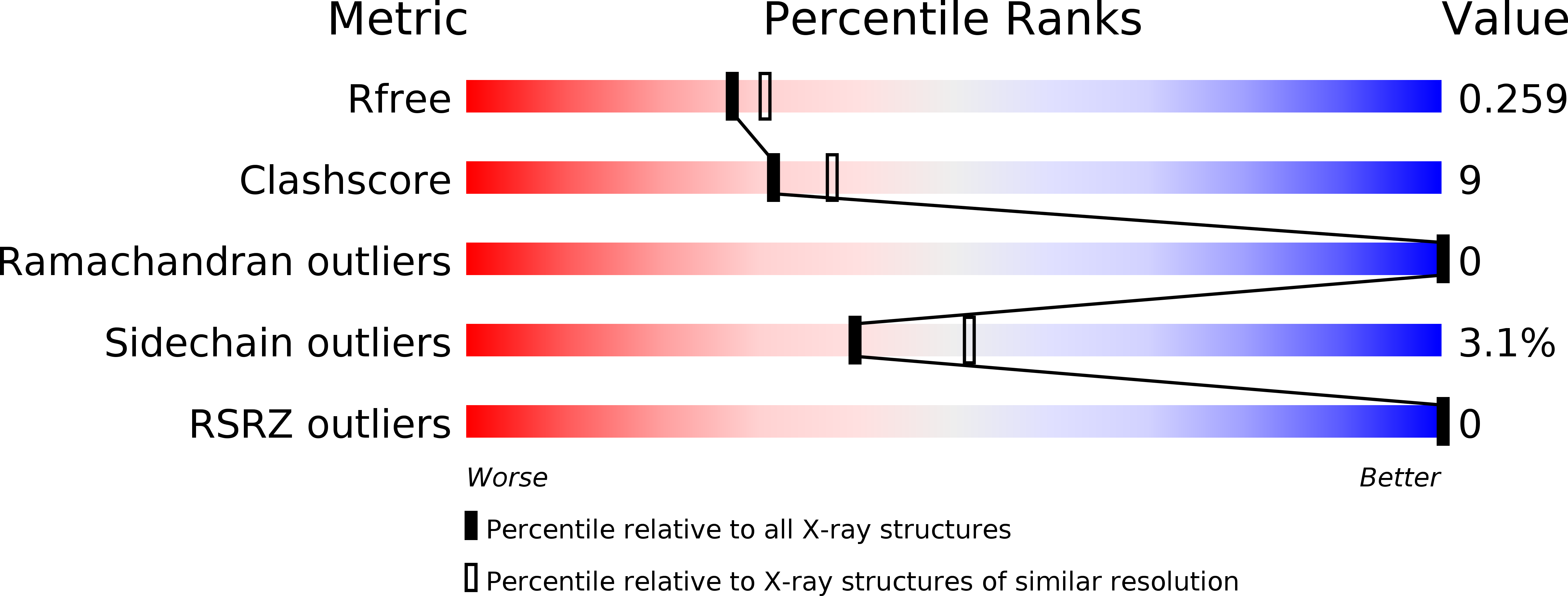
Resolution:
2.20 Å
R-Value Free:
0.26
R-Value Work:
0.19
R-Value Observed:
0.19
Space Group:
P 65 2 2
Macromolecular Entities
Structures with similar UniProt ID
Protein Blast
Polymer Type:polypeptide(L)
Molecule:SCAFFOLDING PROTEIN
Gene (Uniprot):cipC
Chain IDs:A
Chain Length:160
Number of Molecules:1
Biological Source:Clostridium cellulolyticum
Primary Citation
Structure of a family IIIa scaffoldin CBD from the cellulosome of Clostridium cellulolyticum at 2.2 A resolution.
Acta Crystallogr.,Sect.D 56 1560 1568 (2000)
PMID: 11092922 DOI: 10.1107/S0907444900012889

Abstact

The crystal structure of the family IIIa cellulose-binding domain (CBD) from the cellulosomal scaffoldin subunit (CipC) of Clostridium cellulolyticum has been determined. The structure reveals a nine-stranded jelly-roll topology which exhibits distinctive structural elements consistent with family III CBDs that bind crystalline cellulose. These include a well conserved calcium-binding site, a putative cellulose-binding surface and a conserved shallow groove of unknown function. The CipC CBD structure is very similar to the previously elucidated family IIIa CBD from the CipA scaffoldin of C. thermocellum, with some minor differences. The CipC CBD structure was also compared with other previously described CBD structures from families IIIc and IV derived from the endoglucanases of Thermomonospora fusca and Cellulomonas fimi, respectively. The possible functional consequences of structural similarities and differences in the shallow groove and cellulose-binding faces among various CBD families and subfamilies are discussed.

Legend

Protein

Chemical

Disease

Primary Citation of related structures