
Deposition Date
2000-10-25
Release Date
2001-04-21
Last Version Date
2024-02-07
Entry Detail
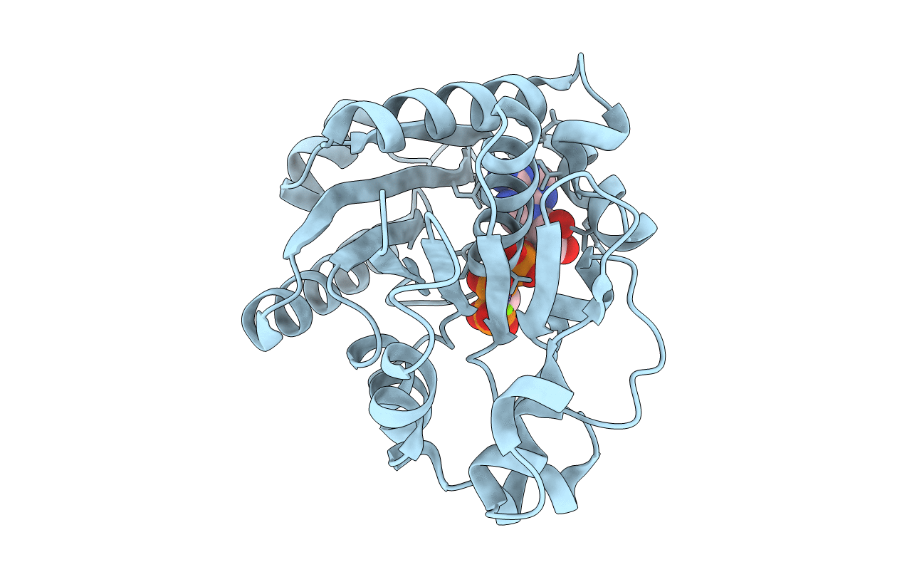
PDB ID:
1G3R
Keywords:
Title:
CRYSTAL STRUCTURE ANALYSIS OF PYROCOCCUS FURIOSUS CELL DIVISION ATPASE MIND
Biological Source:
Source Organism(s):
Pyrococcus furiosus (Taxon ID: 2261)
Expression System(s):
Method Details:
Experimental Method:
Resolution:
2.70 Å
R-Value Free:
0.25
R-Value Work:
0.20
R-Value Observed:
0.20
Space Group:
P 21 3
