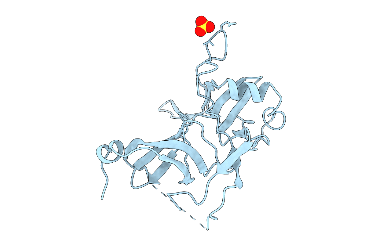
1G3P image
Entry Detail
PDB ID:
1G3P
Keywords:
Title:
CRYSTAL STRUCTURE OF THE N-TERMINAL DOMAINS OF BACTERIOPHAGE MINOR COAT PROTEIN G3P
Biological Source:
Source Organism:
Host Organism:
PDB Version:
Deposition Date:
1997-12-22
Release Date:
1998-01-28
Method Details:
Experimental Method:
Resolution:
1.46 Å
R-Value Free:
0.22
R-Value Work:
0.18
R-Value Observed:
0.18
Space Group:
P 32 2 1
Macromolecular Entities
Polymer Type:polypeptide(L)
Description:MINOR COAT PROTEIN
Chain IDs:A
Chain Length:217
Number of Molecules:1
Biological Source:Enterobacteria phage M13
Modified Residue
Compound ID Chain ID Parent Comp ID Details 2D Image
TRO A TRP 2-HYDROXY-TRYPTOPHAN
Ligand Molecules
Primary Citation
The structural basis of phage display elucidated by the crystal structure of the N-terminal domains of g3p.
Nat.Struct.Biol. 5 140 147 (1998)
PMID: 9461080 DOI: 10.1038/nsb0298-140

Abstact

The structure of the two N-terminal domains of the gene 3 protein of filamentous phages (residues 1-217) has been solved by multiwavelength anomalous diffraction and refined at 1.46 A resolution. Each domain consists of either five or eight beta-strands and a single alpha-helix. Despite missing sequence homology, their cores superimposed with a root-mean-square deviation of 2 A. The domains are engaged in extensive interactions, resulting in a horseshoe shape with aliphatic amino acids and threonines lining the inside, delineating the likely binding site for the F-pilus. The glycine-rich linker connecting the domains is invisible in the otherwise highly ordered structure and may confer flexibility between the domains required during the infection process.

Legend

Protein

Chemical

Disease

Primary Citation of related structures