Deposition Date
2000-10-24
Release Date
2000-11-22
Last Version Date
2024-04-03
Entry Detail
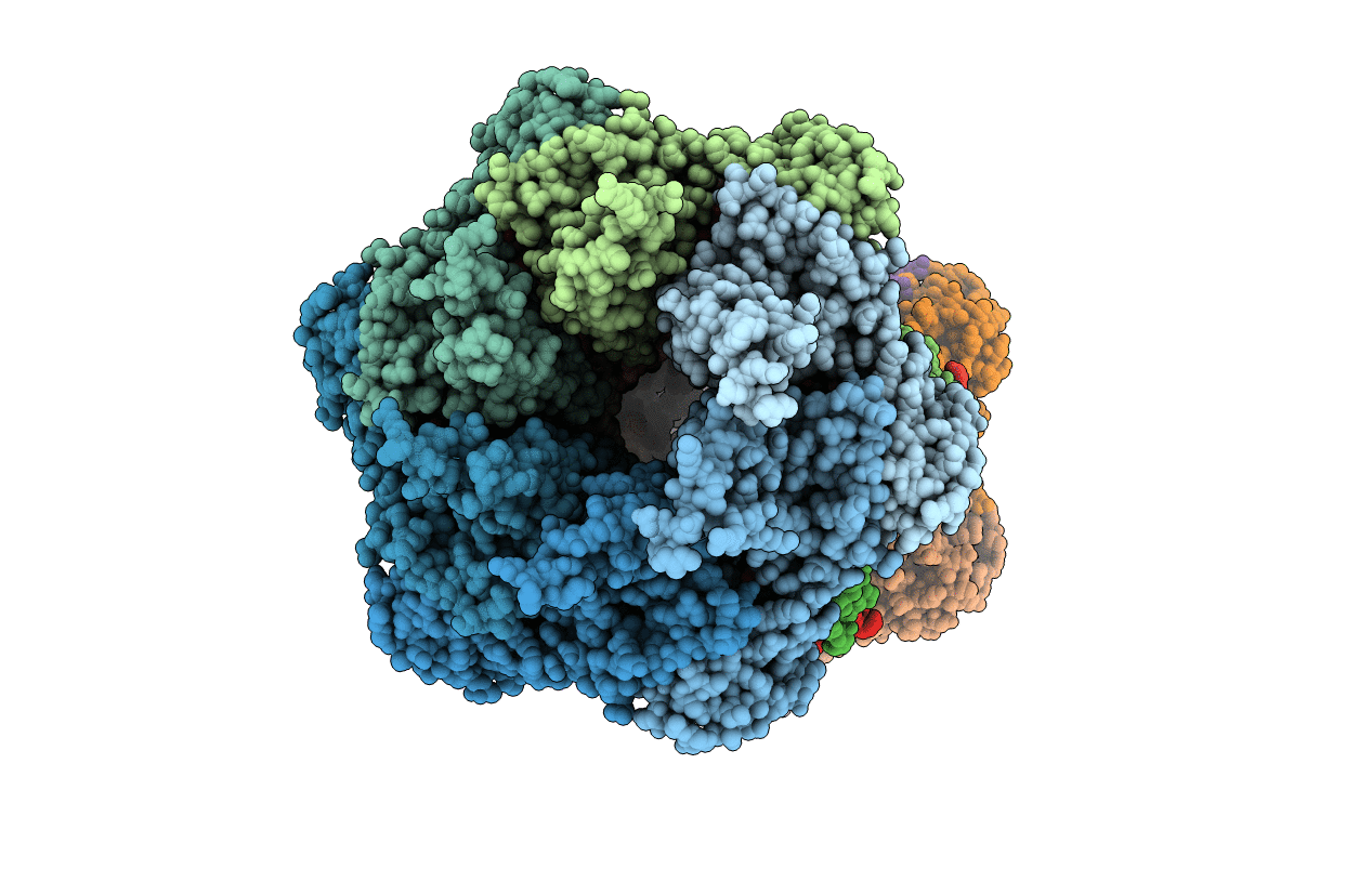
PDB ID:
1G3I
Keywords:
Title:
CRYSTAL STRUCTURE OF THE HSLUV PROTEASE-CHAPERONE COMPLEX
Biological Source:
Source Organism(s):
Haemophilus influenzae (Taxon ID: 727)
Expression System(s):
Method Details:
Experimental Method:
Resolution:
3.41 Å
R-Value Free:
0.28
R-Value Work:
0.24
R-Value Observed:
0.24
Space Group:
P 21 21 2