1G2X image
Deposition Date 2000-10-22
Release Date 2003-06-17
Last Version Date 2024-10-30
Entry Detail
PDB ID:
1G2X
Keywords:
Title:
Sequence induced trimerization of krait PLA2: crystal structure of the trimeric form of krait PLA2
Biological Source:
Source Organism:
Method Details:
Experimental Method:
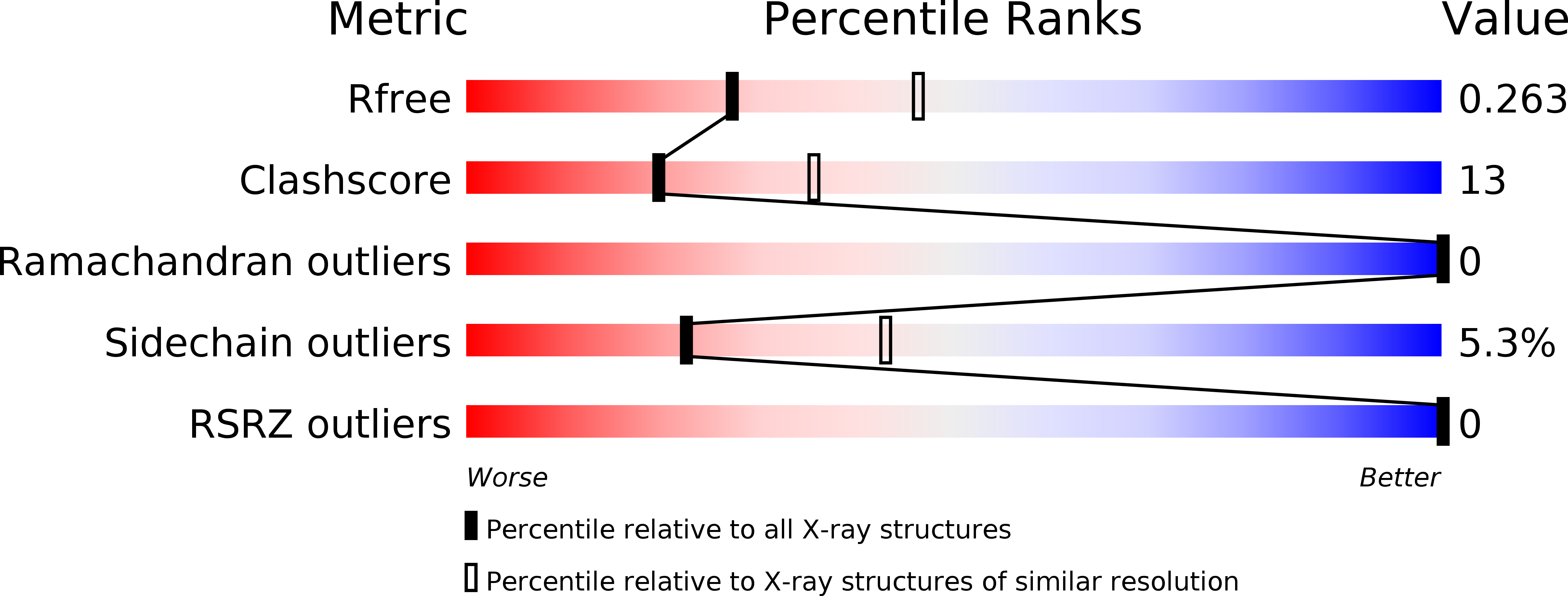
Resolution:
2.50 Å
R-Value Free:
0.26
R-Value Work:
0.19
R-Value Observed:
0.19
Space Group:
C 1 2 1
Macromolecular Entities
Structures with similar UniProt ID
Protein Blast
Polymer Type:polypeptide(L)
Molecule:PHOSPHOLIPASE A2
Chain IDs:A, B, C
Chain Length:118
Number of Molecules:3
Biological Source:Bungarus caeruleus
Primary Citation
Sequence-induced trimerization of phospholipase A2: structure of a trimeric isoform of PLA2 from common krait (Bungarus caeruleus) at 2.5 A resolution.
Acta Crystallogr.,Sect.F 61 8 13 (2005)
PMID: 16508078 DOI: 10.1107/S1744309104025503

Abstact

The venom of the common Indian krait (Bungarus caeruleus) contains about a dozen isoforms of phospholipase A2 (PLA2), which exist in different oligomeric forms as well as in complexes with low-molecular-weight ligands. The basic objective of multimerization and complexation is either to inactivate PLA2 in the venom for long-term storage, to generate a new PLA2 function or to make a more lethal assembly. The current isoform was isolated from the venom of B. caeruleus. Dynamic light-scattering studies indicated the presence of a stable trimeric association of this PLA2. Its primary sequence was determined by cDNA cloning. The purified protein was crystallized with 2.8 M NaCl as a precipitating agent using the sitting-drop vapour-diffusion method. The crystals belonged to the monoclinic space group C2, with unit-cell parameters a = 80.9, b = 80.5, c = 57.1 A, beta = 90.3 degrees. The structure was refined to a final R factor of 0.198. This is a novel trimeric PLA2 structure in which the central pore formed by the association of three molecules is filled with water molecules. The interactions across the pore take place via multiple water bridges primarily to the side chains of Arg, Lys and Thr residues. Approximately 12% of the total solvent-accessible surface area is buried in the core of the trimer. The active sites of all three molecules are located on the surface and are fully exposed to the solvent, resulting in a highly potent enzymatic unit.

Legend

Protein

Chemical

Disease

Primary Citation of related structures