Deposition Date
2000-10-20
Release Date
2001-04-21
Last Version Date
2024-02-07
Entry Detail
PDB ID:
1G2N
Keywords:
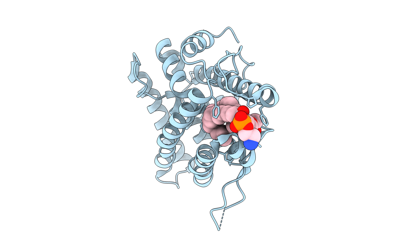
Title:
CRYSTAL STRUCTURE OF THE LIGAND BINDING DOMAIN OF THE ULTRASPIRACLE PROTEIN USP, THE ORTHOLOG OF RXRS IN INSECTS
Biological Source:
Source Organism(s):
Heliothis virescens (Taxon ID: 7102)
Expression System(s):
Method Details:
Experimental Method:
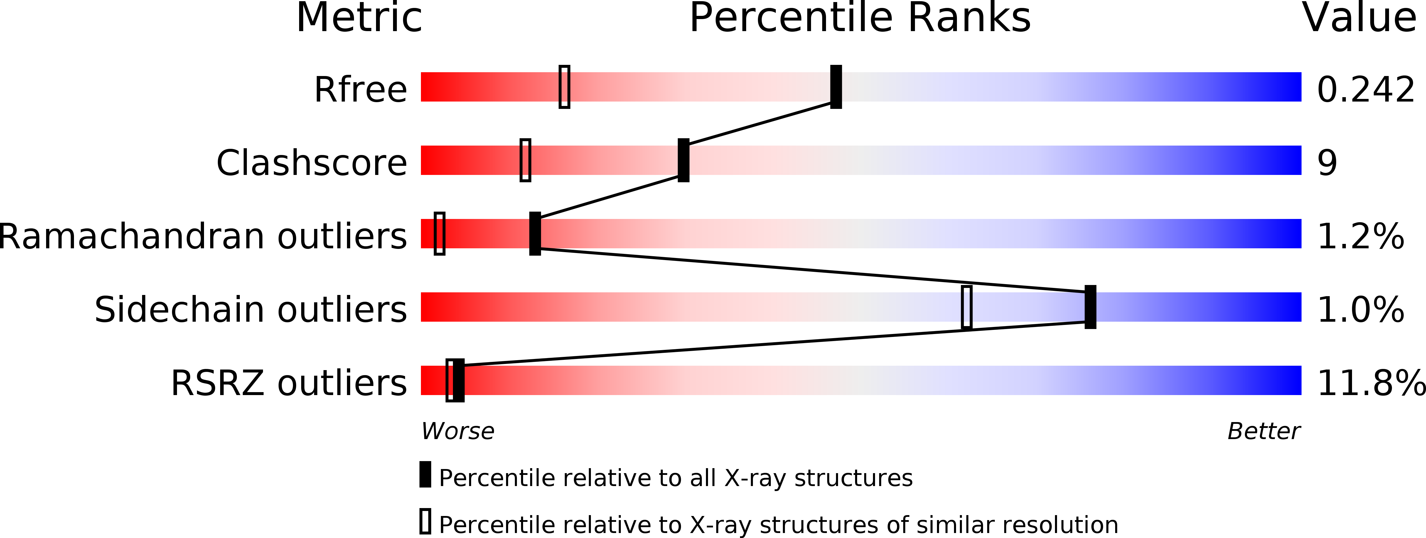
Resolution:
1.65 Å
R-Value Free:
0.24
R-Value Work:
0.21
R-Value Observed:
0.21
Space Group:
P 43 2 2