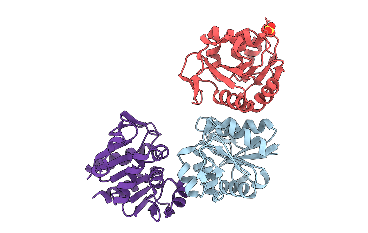
1G2I image
Deposition Date 2000-10-19
Release Date 2000-11-08
Last Version Date 2024-10-16
Entry Detail
PDB ID:
1G2I
Keywords:
Title:
CRYSTAL STRUCTURE OF A NOVEL INTRACELLULAR PROTEASE FROM PYROCOCCUS HORIKOSHII AT 2 A RESOLUTION
Biological Source:
Source Organism:
Host Organism:
Method Details:
Experimental Method:
Resolution:
2.00 Å
R-Value Free:
0.2
R-Value Work:
0.18
R-Value Observed:
0.18
Space Group:
P 41 21 2
Macromolecular Entities
Structures with similar UniProt ID
Protein Blast
Polymer Type:polypeptide(L)
Molecule:PROTEASE I
Gene (Uniprot):PH1704
Chain IDs:A, B, C
Chain Length:166
Number of Molecules:3
Biological Source:Pyrococcus horikoshii
Modified Residue
Compound ID Chain ID Parent Comp ID Details 2D Image
MSE A MET SELENOMETHIONINE
Ligand Molecules
Primary Citation
Crystal structure of an intracellular protease from Pyrococcus horikoshii at 2-A resolution.
Proc.Natl.Acad.Sci.USA 97 14079 14084 (2000)
PMID: 11114201 DOI: 10.1073/pnas.260503597

Abstact

The intracellular protease from Pyrococcus horikoshii (PH1704) and PfpI from Pyrococcus furiosus are members of a class of intracellular proteases that have no sequence homology to any other known protease family. We report the crystal structure of PH1704 at 2.0-A resolution. The protease is tentatively identified as a cysteine protease based on the presence of cysteine (residue 100) in a nucleophile elbow motif. In the crystal, PH1704 forms a hexameric ring structure, and the active sites are formed at the interfaces between three pairs of monomers.

Legend

Protein

Chemical

Disease

Primary Citation of related structures