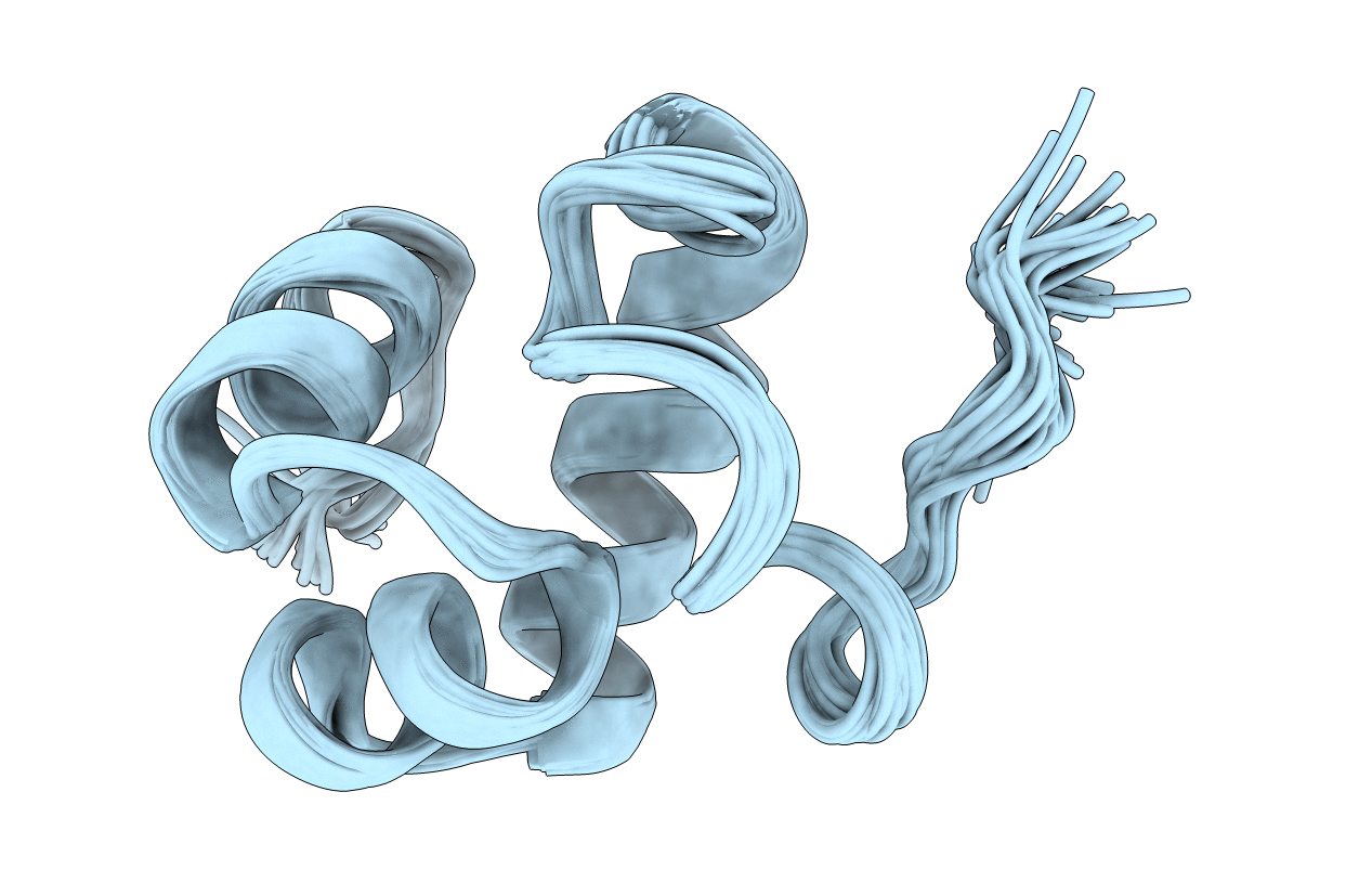
1G2H image
Deposition Date 2000-10-19
Release Date 2001-03-07
Last Version Date 2024-05-22
Entry Detail
PDB ID:
1G2H
Keywords:
Title:
SOLUTION STRUCTURE OF THE DNA-BINDING DOMAIN OF THE TYRR PROTEIN OF HAEMOPHILUS INFLUENZAE
Biological Source:
Source Organism:
Host Organism:
Method Details:
Experimental Method:
Conformers Calculated:
100
Conformers Submitted:
32
Selection Criteria:
structures with acceptable covalent geometry,structures with favorable non-bond energy,structures with the least restraint violations,structures with the lowest energy
Macromolecular Entities
Structures with similar UniProt ID
Protein Blast
Polymer Type:polypeptide(L)
Molecule:TRANSCRIPTIONAL REGULATORY PROTEIN TYRR HOMOLOG
Gene (Uniprot):tyrR
Chain IDs:A
Chain Length:61
Number of Molecules:1
Biological Source:Haemophilus influenzae
Ligand Molecules
Primary Citation
Solution structure of the DNA-binding domain of the TyrR protein of Haemophilus influenzae.
Protein Sci. 10 592 598 (2001)
PMID: 11344327 DOI: 10.1110/ps.45301

Abstact

The TyrR protein of Haemophilus influenzae is a 36-kD transcription factor whose major function is to control the expression of genes important in the biosynthesis and transport of aromatic amino acids. Using (1)H and (15)N NMR spectroscopy, we have determined the 3D solution structure of the TyrR C-terminal DNA-binding domain (DBD) containing residues from 258 to 318 (TyrR[258-318]). The NMR results show that this segment of TyrR consists of a potential hinge helix at its N terminus (residues 263-270) as well as three well-defined alpha-helices extending from residues 277-289 (HR-2), 293-300 (HR-1), and 304-314 (HR). Helix HR-1 and HR fold in a typical helix-turn-helix (HTH) motif. The three helices and the hinge helix are tightly bound together by hydrophobic interaction and hydrogen bonds. Several hydrophilic residues whose side chains may directly interact with DNA are identified. A hydrophobic patch that may be part of the interaction surface between the domains of TyrR protein is also observed. Comparisons with the structures of other HTH DNA-binding proteins reveal that in terms of the spatial orientation of the three helices, this protein most closely resembles the cap family.

Legend

Protein

Chemical

Disease

Primary Citation of related structures