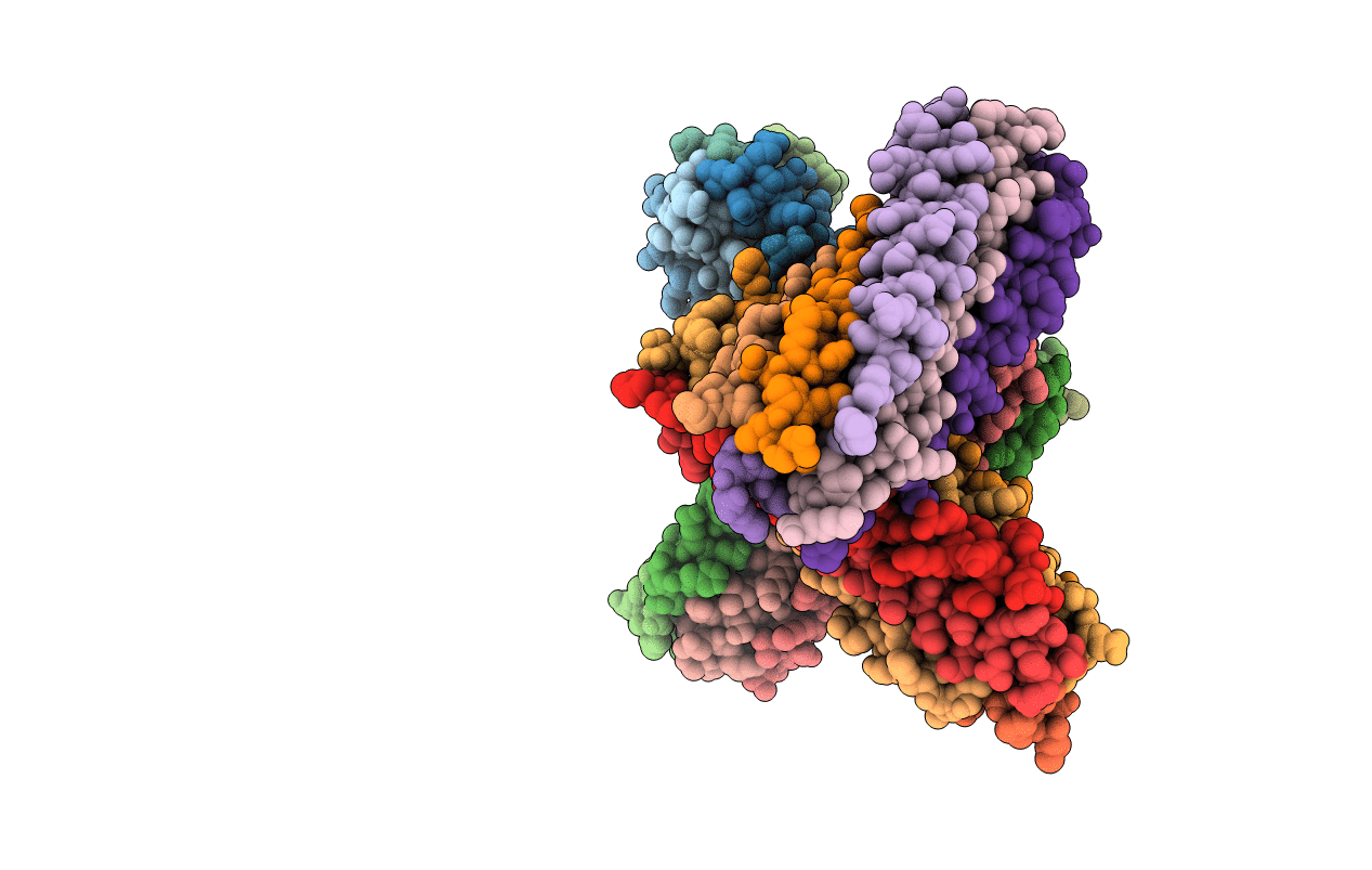
1G2C image
Deposition Date 2000-10-18
Release Date 2001-01-03
Last Version Date 2024-02-07
Entry Detail
PDB ID:
1G2C
Keywords:
Title:
HUMAN RESPIRATORY SYNCYTIAL VIRUS FUSION PROTEIN CORE
Biological Source:
Source Organism:
Host Organism:
Method Details:
Experimental Method:
Resolution:
2.30 Å
R-Value Free:
0.28
R-Value Work:
0.23
R-Value Observed:
0.23
Space Group:
P 1
Macromolecular Entities
Structures with similar UniProt ID
Protein Blast
Polymer Type:polypeptide(L)
Molecule:FUSION PROTEIN (F)
Gene (Uniprot):F
Chain IDs:A, C, E, G, I, K, M, O, Q, S, U, W
Chain Length:52
Number of Molecules:12
Biological Source:Human respiratory syncytial virus
Structures with similar UniProt ID
Protein Blast
Polymer Type:polypeptide(L)
Molecule:FUSION PROTEIN (F)
Gene (Uniprot):F
Chain IDs:B, D, F, H, J, L, N, P, R, T, V, X
Chain Length:43
Number of Molecules:12
Biological Source:Human respiratory syncytial virus
Primary Citation
Structural characterization of the human respiratory syncytial virus fusion protein core.
Proc.Natl.Acad.Sci.USA 97 14172 14177 (2000)
PMID: 11106388 DOI: 10.1073/pnas.260499197

Abstact

Human respiratory syncytial virus (HRSV) is a major cause of a number of severe respiratory diseases, including bronchiolitis and pneumonia, in infants and young children. The HRSV F protein, a glycoprotein essential for viral entry, is a primary target for vaccine and drug development. Two heptad-repeat regions within the HRSV F sequence were predicted by the computer program learncoil-vmf. These regions are thought to form trimer-of-hairpins-like structures, similar to those found in the fusion proteins of several enveloped viruses. The hairpin structure likely brings the viral and cellular membranes into close apposition, thereby facilitating membrane fusion and subsequent viral entry. Here, we show that peptides, denoted HR-N and HR-C, corresponding to the heptad-repeat regions from the N-terminal and C-terminal segments of the HRSV F protein, respectively, form a stable alpha-helical trimer of heterodimers. The HRSV N/C complex was crystallized and its x-ray structure was determined at 2.3-A resolution. As anticipated, the complex is a six-helix bundle in which the HR-N peptides form a three-stranded, central coiled coil, and the HR-C peptides pack in an antiparallel manner into hydrophobic grooves on the coiled-coil surface. There is remarkable structural similarity between the HRSV N/C complex and the fusion protein core of other viruses, including HIV-1 gp41. In addition, earlier work has shown that HRSV HR-C peptides, like the HIV-1 gp41 C peptides, inhibit viral infection. Thus, drug discovery and vaccine development strategies aimed at inhibiting viral entry by blocking hairpin formation may be applied to the inhibition of HRSV.

Legend

Protein

Chemical

Disease

Primary Citation of related structures
Feedback Form
Name
Email
Institute
Feedback