Deposition Date
2000-10-17
Release Date
2001-03-21
Last Version Date
2024-02-07
Entry Detail
PDB ID:
1G28
Keywords:
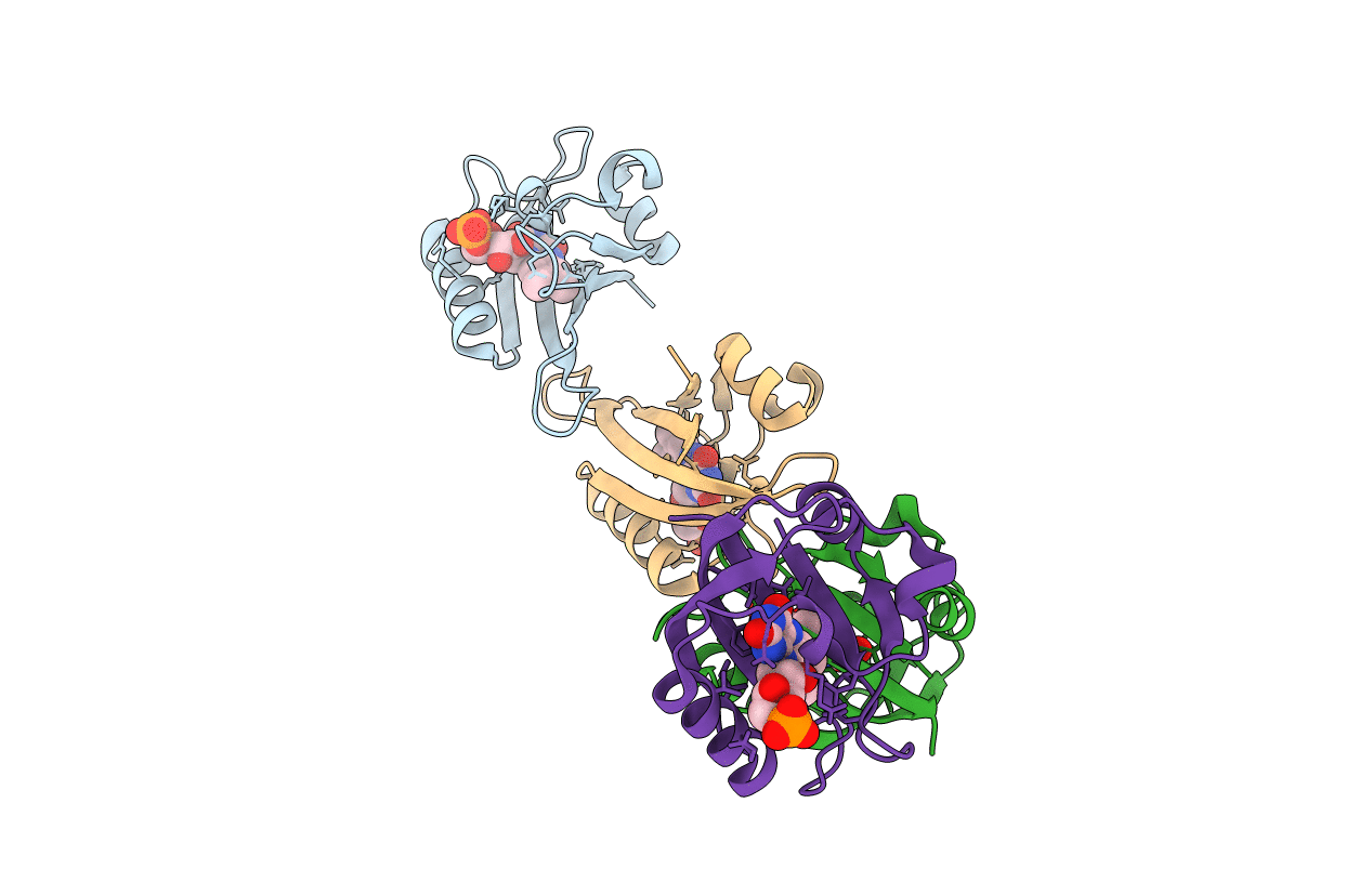
Title:
STRUCTURE OF A FLAVIN-BINDING DOMAIN, LOV2, FROM THE CHIMERIC PHYTOCHROME/PHOTOTROPIN PHOTORECEPTOR PHY3
Biological Source:
Source Organism(s):
Adiantum capillus-veneris (Taxon ID: 13818)
Expression System(s):
Method Details:
Experimental Method:
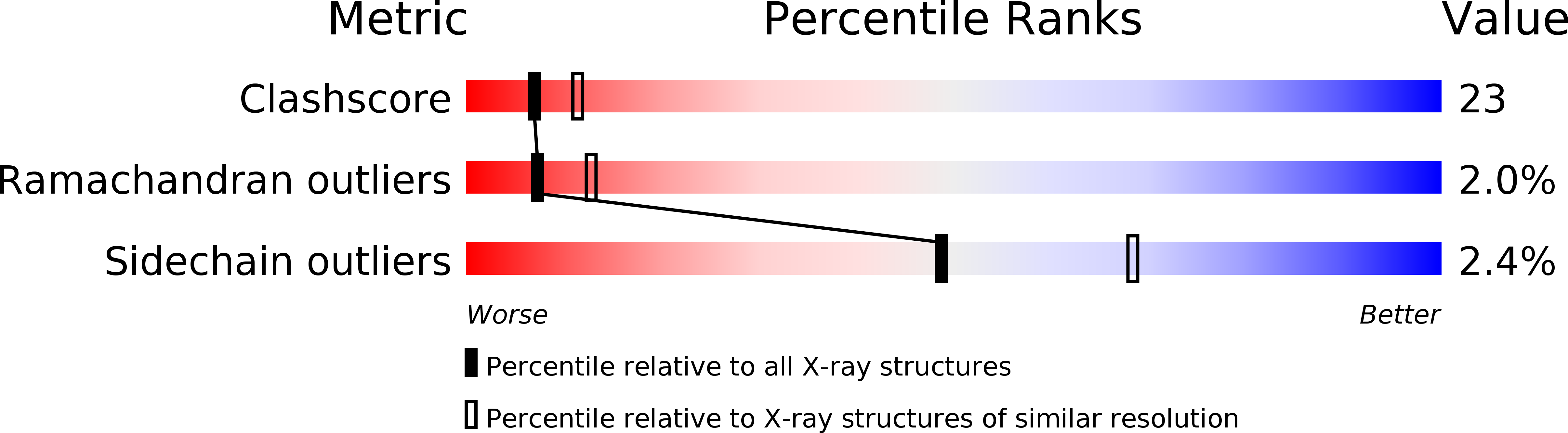
Resolution:
2.73 Å
R-Value Free:
0.27
R-Value Work:
0.24
R-Value Observed:
0.24
Space Group:
P 1