1FZR image
Deposition Date 2000-10-04
Release Date 2001-01-17
Last Version Date 2024-02-07
Entry Detail
PDB ID:
1FZR
Keywords:
Title:
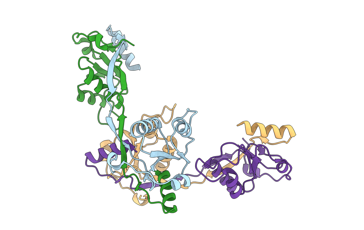
CRYSTAL STRUCTURE OF BACTERIOPHAGE T7 ENDONUCLEASE I
Biological Source:
Source Organism:
Host Organism:
Method Details:
Experimental Method:
Resolution:
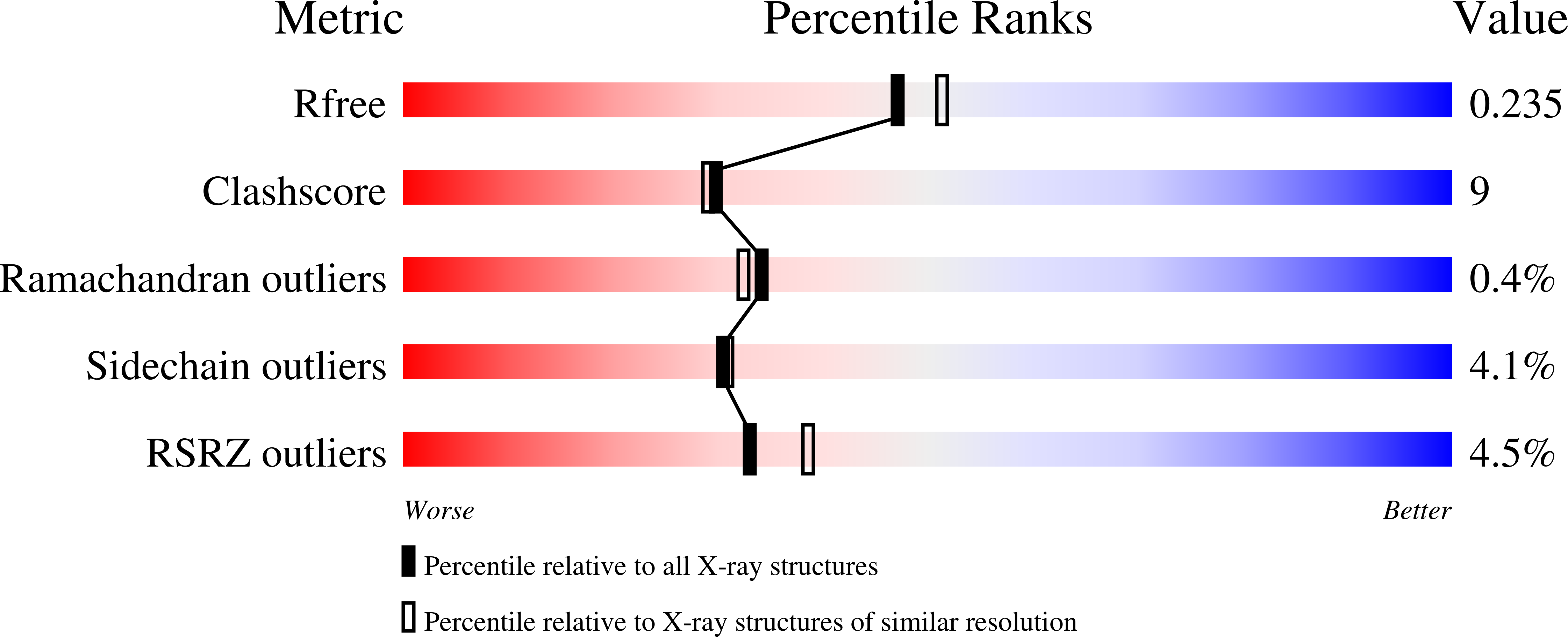
2.10 Å
R-Value Free:
0.23
R-Value Work:
0.19
R-Value Observed:
0.20
Space Group:
P 21 21 2
Macromolecular Entities
Polymer Type:polypeptide(L)
Molecule:ENDONUCLEASE I
Gene (Uniprot):3
Mutations:E65K
Chain IDs:A, B, C, D
Chain Length:138
Number of Molecules:4
Biological Source:Enterobacteria phage T7
Primary Citation
Crystal structure of the Holliday junction resolving enzyme T7 endonuclease I.
Nat.Struct.Biol. 8 62 67 (2001)
PMID: 11135673 DOI: 10.1038/83067

Abstact

We have solved the crystal structure of the Holliday junction resolving enzyme T7 endonuclease I at 2.1 A resolution using the multiwavelength anomalous dispersion (MAD) technique. Endonuclease I exhibits strong structural specificity for four-way DNA junctions. The structure shows that it forms a symmetric homodimer arranged in two well-separated domains. Each domain, however, is composed of elements from both subunits, and amino acid side chains from both protomers contribute to the active site. While no significant structural similarity could be detected with any other junction resolving enzyme, the active site is similar to that found in several restriction endonucleases. T7 endonuclease I therefore represents the first crystal structure of a junction resolving enzyme that is a member of the nuclease superfamily of enzymes.

Legend

Protein

Chemical

Disease

Primary Citation of related structures