1FXJ image
Deposition Date 2000-09-26
Release Date 2000-10-18
Last Version Date 2024-10-09
Entry Detail
PDB ID:
1FXJ
Keywords:
Title:
CRYSTAL STRUCTURE OF N-ACETYLGLUCOSAMINE 1-PHOSPHATE URIDYLTRANSFERASE
Biological Source:
Source Organism:
Escherichia coli (Taxon ID: 562)
Host Organism:
Method Details:
Experimental Method:
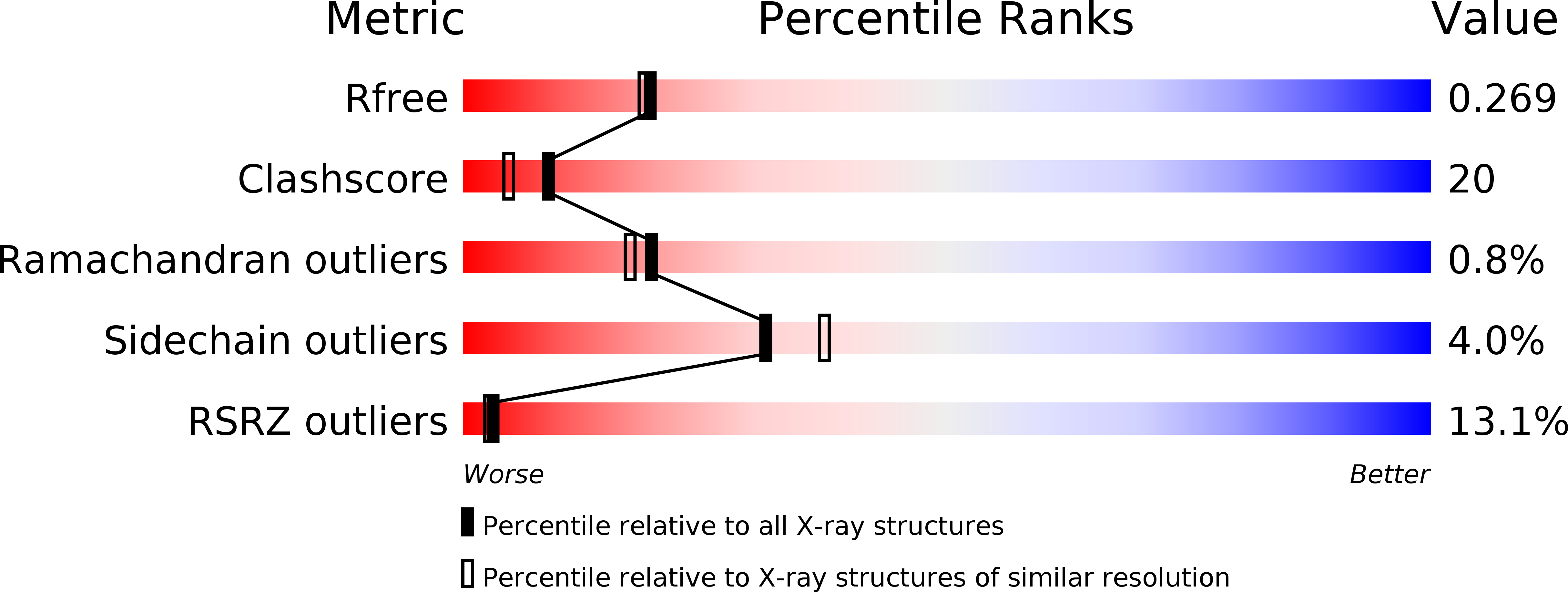
Resolution:
2.25 Å
R-Value Free:
0.27
R-Value Work:
0.23
Space Group:
H 3 2
Macromolecular Entities
Polymer Type:polypeptide(L)
Molecule:UDP-N-ACETYLGLUCOSAMINE PYROPHOSPHORYLASE
Gene (Uniprot):glmU
Chain IDs:A, B
Chain Length:331
Number of Molecules:2
Biological Source:Escherichia coli
Primary Citation
Crystal structure of the bifunctional N-acetylglucosamine 1-phosphate uridyltransferase from Escherichia coli: a paradigm for the related pyrophosphorylase superfamily.
EMBO J. 18 4096 4107 (1999)
PMID: 10428949 DOI: 10.1093/emboj/18.15.4096

Abstact

N-acetylglucosamine 1-phosphate uridyltransferase (GlmU) is a cytoplasmic bifunctional enzyme involved in the biosynthesis of the nucleotide-activated UDP-GlcNAc, which is an essential precursor for the biosynthetic pathways of peptidoglycan and other components in bacteria. The crystal structure of a truncated form of GlmU has been solved at 2.25 A resolution using the multiwavelength anomalous dispersion technique and its function tested with mutagenesis studies. The molecule is composed of two distinct domains connected by a long alpha-helical arm: (i) an N-terminal domain which resembles the dinucleotide-binding Rossmann fold; and (ii) a C-terminal domain which adopts a left-handed parallel beta-helix structure (LbetaH) as found in homologous bacterial acetyltransferases. Three GlmU molecules assemble into a trimeric arrangement with tightly packed parallel LbetaH domains, the long alpha-helical linkers being seated on top of the arrangement and the N-terminal domains projected away from the 3-fold axis. In addition, the 2.3 A resolution structure of the GlmU-UDP-GlcNAc complex reveals the structural bases required for the uridyltransferase activity. These structures exemplify a three-dimensional template for the development of new antibacterial agents and for studying other members of the large family of XDP-sugar bacterial pyrophosphorylases.

Legend

Protein

Chemical

Disease

Primary Citation of related structures