1FXA image
Entry Detail
PDB ID:
1FXA
Title:
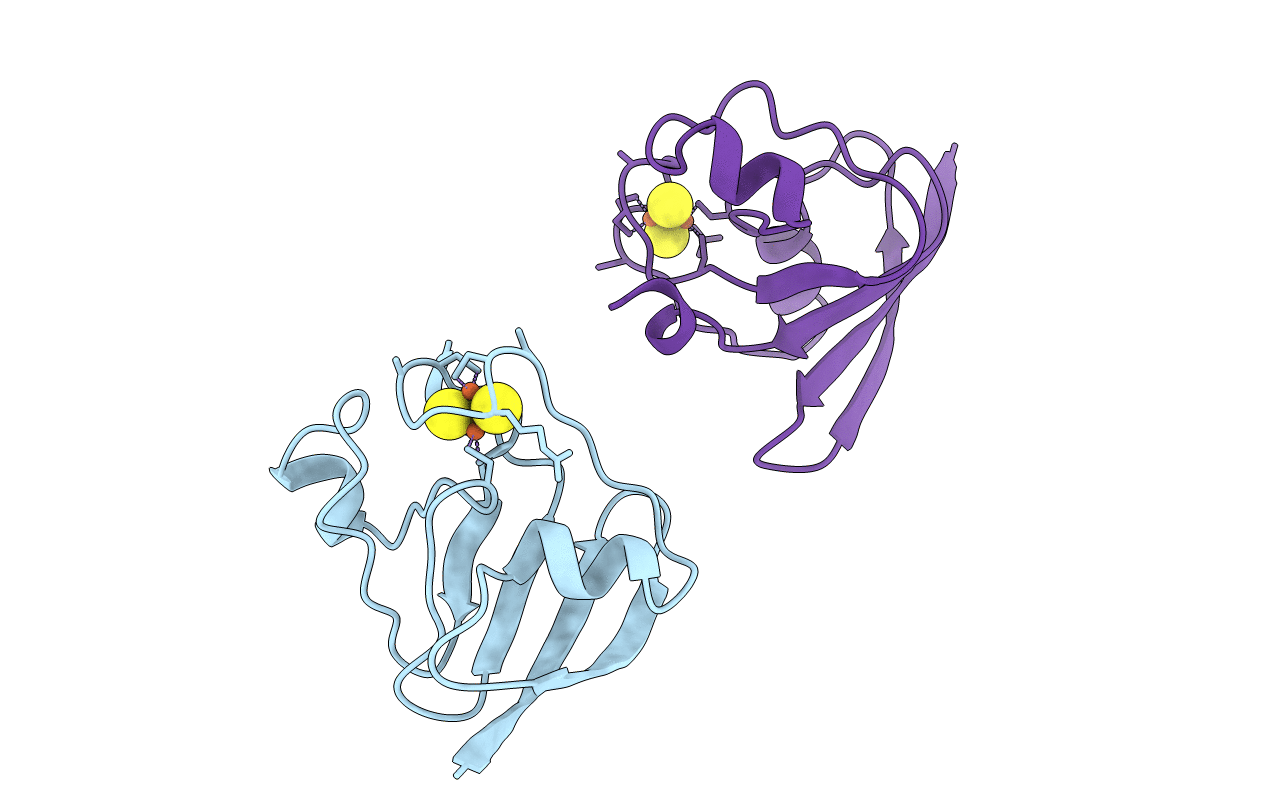
CRYSTALLIZATION AND STRUCTURE DETERMINATION TO 2.5-ANGSTROMS RESOLUTION OF THE OXIDIZED [2FE-2S] FERREDOXIN ISOLATED FROM ANABAENA 7120
Biological Source:
Source Organism:
PDB Version:
Deposition Date:
1991-01-09
Release Date:
1992-07-15
Method Details:
Experimental Method:
Resolution:
2.50 Å
R-Value Observed:
0.18
Space Group:
P 21 21 21
Macromolecular Entities
Polymer Type:polypeptide(L)
Description:[2FE-2S] FERREDOXIN
Chain IDs:A, B
Chain Length:98
Number of Molecules:2
Biological Source:Nostoc sp.
Ligand Molecules
Primary Citation
Crystallization and structure determination to 2.5-A resolution of the oxidized [2Fe-2S] ferredoxin isolated from Anabaena 7120.
Biochemistry 30 4126 4131 (1991)
PMID: 1902376 DOI: 10.1021/bi00231a003

Abstact

The molecular structure of the oxidized form of the [2Fe-2S] ferredoxin isolated from the cyanobacterium Anabaena species strain PCC 7120 has been determined by X-ray diffraction analysis to a nominal resolution of 2.5 A and refined to a crystallographic R factor of 18.7%. Crystals used in this investigation belong to the space group P2(1)2(1)2(1) with unit cell dimensions of a = 37.42 A, b = 38.12 A, and c = 147.12 A and two molecules in the asymmetric unit. The three-dimensional structure of this ferredoxin was solved by a method that combined X-ray data from one isomorphous heavy-atom derivative with noncrystallographic symmetry averaging and solvent flattening. As in other plant-type [2Fe-2S] ferredoxins, the iron-sulfur cluster is located toward the outer edge of the molecule, and the irons are tetrahedrally coordinated by both inorganic sulfurs and sulfurs provided by protein cysteine residues. The main secondary structural elements include four strands of beta-pleated sheet and three alpha-helical regions.

Legend

Protein

Chemical

Disease

Primary Citation of related structures