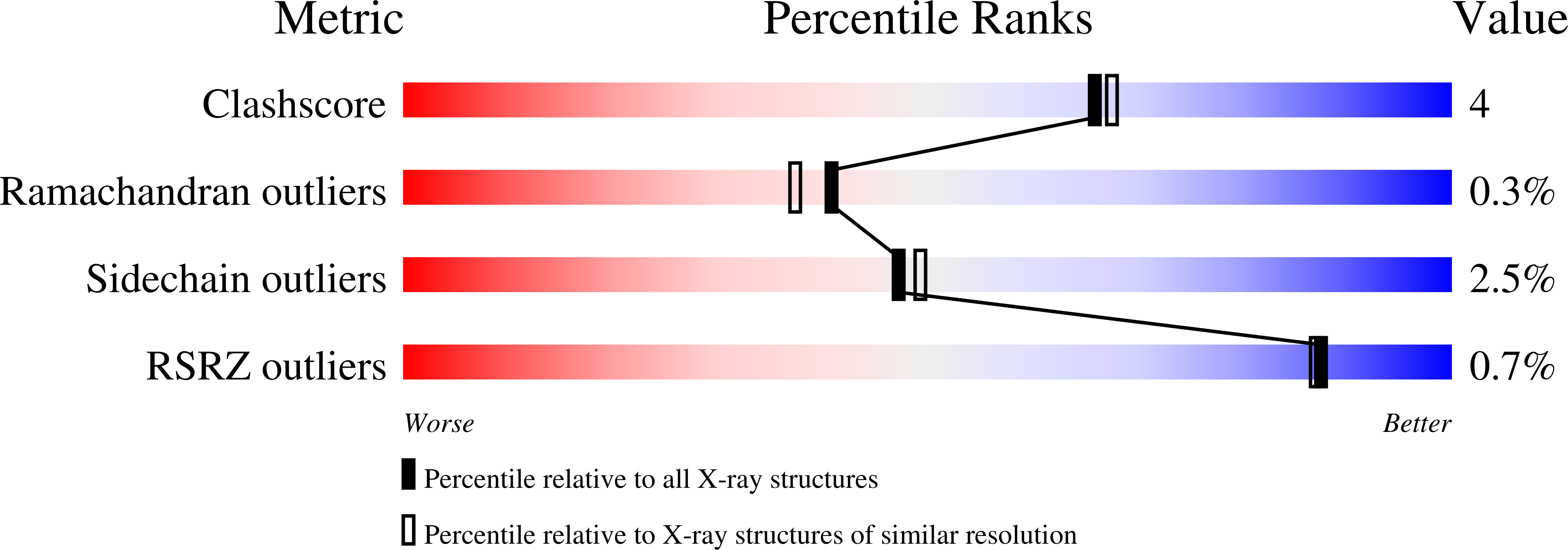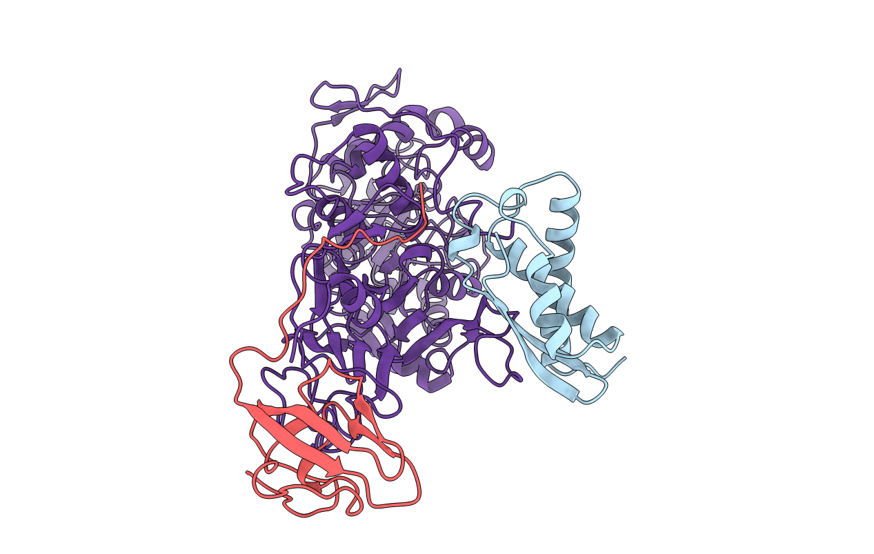
1FWI image
Deposition Date 1997-04-23
Release Date 1997-10-15
Last Version Date 2021-11-03
Entry Detail
PDB ID:
1FWI
Keywords:
Title:
KLEBSIELLA AEROGENES UREASE, H134A VARIANT
Biological Source:
Source Organism:
Host Organism:
Method Details:
Experimental Method:
Resolution:
2.00 Å
R-Value Work:
0.17
R-Value Observed:
0.17
Space Group:
I 21 3
Macromolecular Entities
Polymer Type:polypeptide(L)
Molecule:UREASE
Gene (Uniprot):ureA
Mutations:H(C 134)A
Chain IDs:A
Chain Length:100
Number of Molecules:1
Biological Source:Klebsiella aerogenes
Polymer Type:polypeptide(L)
Molecule:UREASE
Gene (Uniprot):ureB
Mutations:H(C 134)A
Chain IDs:B
Chain Length:106
Number of Molecules:1
Biological Source:Klebsiella aerogenes
Polymer Type:polypeptide(L)
Molecule:UREASE
Gene (Uniprot):ureC
Mutations:H(C 134)A
Chain IDs:C
Chain Length:567
Number of Molecules:1
Biological Source:Klebsiella aerogenes
Modified Residue
Compound ID Chain ID Parent Comp ID Details 2D Image
KCX C LYS LYSINE NZ-CARBOXYLIC ACID
Ligand Molecules
Primary Citation
Characterization of the mononickel metallocenter in H134A mutant urease.
J.Biol.Chem. 271 18632 18637 (1996)
PMID: 8702515 DOI: 10.1074/jbc.271.31.18632

Abstact

A mutant form of Klebsiella aerogenes urease possessing Ala instead of His at position 134 (H134A) is inactive and binds approximately half the normal complement of nickel (Park, I.-S., and Hausinger, R. P.(1993) Protein Sci. 2, 1034-1041). The crystal structure of the H134A protein was obtained at 2.0-A resolution, and it confirms that only Ni-1 of the two nickel ions found in the native enzyme is present. In contrast to the pseudotetrahedral geometry observed for Ni-1 in native urease (where it is liganded by His-246, His-272, one oxygen atom of carbamylated Lys-217, and a water molecule at partial occupancy), the mononickel metallocenter in the H134A protein was found to possess octahedral geometry and was coordinated by the above protein ligands plus three water molecules. The nickel site of H134A urease was probed by UV-visible, variable temperature magnetic circular dichroism, and x-ray absorption spectroscopies. The spectroscopic data are consistent with the presence of Ni(II) in octahedral geometry coordinated by two histidylimidazoles and additional oxygen and/or nitrogen donors. These data underscore the requirement of Ni-2 for formation of active urease and demonstrate the important role of Ni-2 in establishing the proper Ni-1 coordination geometry.

Legend

Protein

Chemical

Disease

Primary Citation of related structures