Deposition Date
2000-09-20
Release Date
2000-11-15
Last Version Date
2024-11-20
Entry Detail
PDB ID:
1FW0
Keywords:
Title:
CRYSTAL STRUCTURE OF THE GLUR2 LIGAND BINDING CORE (S1S2J) IN COMPLEX WITH KAINATE AT 2.0 A RESOLUTION
Biological Source:
Source Organism(s):
Rattus norvegicus (Taxon ID: 10116)
Expression System(s):
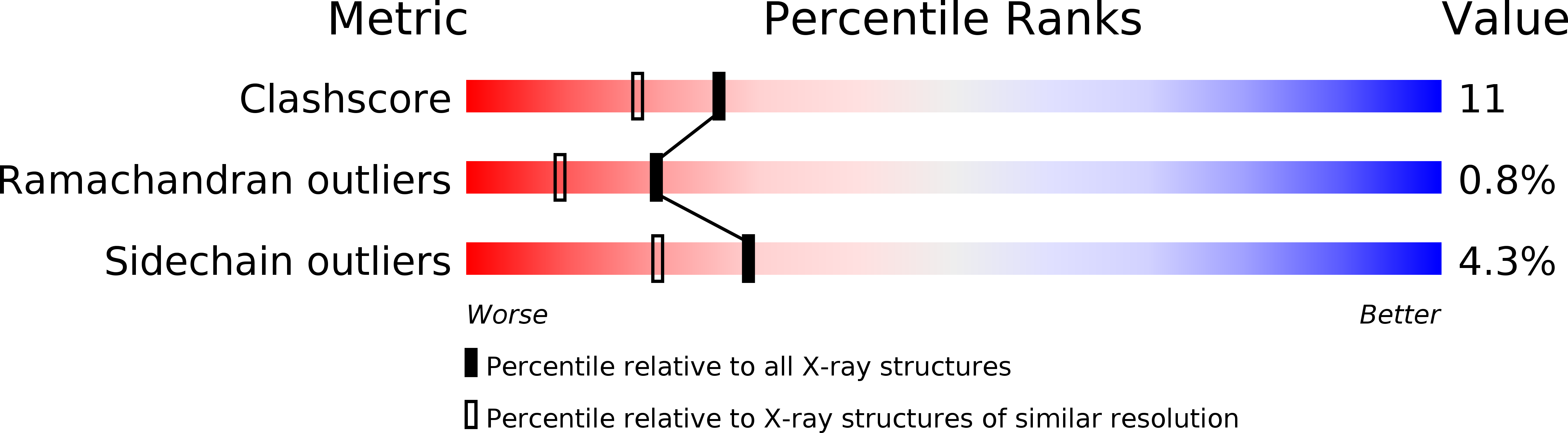
Method Details:
Experimental Method:
Resolution:
1.90 Å
R-Value Free:
0.30
R-Value Work:
0.24
R-Value Observed:
0.28
Space Group:
P 21 21 2