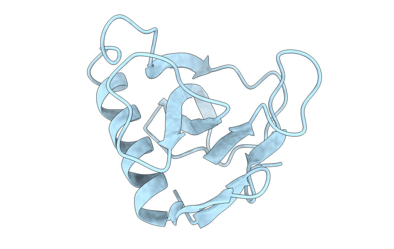
1FUS image
Deposition Date 1993-01-18
Release Date 1993-10-31
Last Version Date 2024-11-06
Entry Detail
PDB ID:
1FUS
Title:
CRYSTAL STRUCTURES OF RIBONUCLEASE F1 OF FUSARIUM MONILIFORME IN ITS FREE FORM AND IN COMPLEX WITH 2'GMP
Biological Source:
Source Organism:
Method Details:
Experimental Method:
Resolution:
1.30 Å
R-Value Observed:
0.18
Space Group:
P 21 21 21
Macromolecular Entities
Polymer Type:polypeptide(L)
Molecule:RIBONUCLEASE F1
Chain IDs:A
Chain Length:106
Number of Molecules:1
Biological Source:Gibberella fujikuroi
Modified Residue
Compound ID Chain ID Parent Comp ID Details 2D Image
PCA A GLN PYROGLUTAMIC ACID
Primary Citation
Crystal structures of ribonuclease F1 of Fusarium moniliforme in its free form and in complex with 2'GMP.
J.Mol.Biol. 230 979 996 (1993)
PMID: 8386773 DOI: 10.1006/jmbi.1993.1214

Abstact

RNase F1, a guanine-specific ribonuclease from Fusarium moniliforme, was crystallized in two different forms, in the absence of an inhibitor and in the presence of 2'GMP. The crystal structure of the RNase F1 free form was solved by the molecular replacement method, using the co-ordinates of the RNase T1 complex with 2'GMP, and was refined to a final R-factor of 18.7%, using the data extended to 1.3 A resolution. For the crystal structure of the RNase F1 complex with 2'GMP, the solution of the molecular replacement method was obtained on the basis of the co-ordinates of the RNase F1 free form, and was refined to a final R-factor of 16.8%, using the data up to 2 A resolution. The two crystal structures of the RNase F1 free form and the complex with 2'GMP are very similar to each other as reflected by a small root-mean-square displacement (r.m.s.d.) value of 0.43 A for all C alpha atoms. The main differences between the two structures are associated with binding of 2'GMP in the substrate recognition site in the loop between Tyr42 and Glu46. A structural comparison between RNase F1 and RNase T1 shows a substantial similarity between all the C alpha atoms, as evidenced by a r.m.s.d. value of 1.4 A. The loop from residues 32 to 38 was strikingly different between these two enzymes, in both its conformation and its hydrogen bonding schemes. The side-chain of a catalytically active residue, His92, is shifted away from the catalytic site in RNase F1 by 1.3 A and 0.85 A with respect to the corresponding positions in the RNase T1 free form and in the RNase T1 complex with 2'GMP, respectively. In the RNase F1 complex, the guanine base of 2'GMP has a syn conformation about the glycosyl bond, and the furanose ring assumes a 3'-exo pucker, which is different from that found in the complex with RNase T1. In the catalytic site of the RNase F1 complex with 2'GMP, one water molecule was observed, which bridges the phosphate oxygen atoms of 2'GMP and the side-chains of the catalytically important residues, His92 and Arg77, through hydrogen bonds. A water molecule occupying the same position was found in the RNase F1 free form. The significance of this water molecule in the hydrolytic reaction is discussed.

Legend

Protein

Chemical

Disease

Primary Citation of related structures