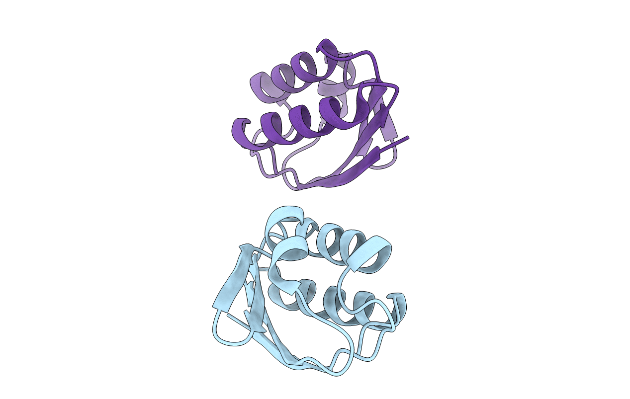
1FU0 image
Deposition Date 2000-09-13
Release Date 2000-11-22
Last Version Date 2024-10-16
Entry Detail
PDB ID:
1FU0
Title:
CRYSTAL STRUCTURE ANALYSIS OF THE PHOSPHO-SERINE 46 HPR FROM ENTEROCOCCUS FAECALIS
Biological Source:
Source Organism:
Method Details:
Experimental Method:
Resolution:
1.90 Å
R-Value Free:
0.23
R-Value Work:
0.17
R-Value Observed:
0.17
Space Group:
P 1 21 1
Macromolecular Entities
Polymer Type:polypeptide(L)
Molecule:PHOSPHOCARRIER PROTEIN HPR
Gene (Uniprot):ptsH
Chain IDs:A, B
Chain Length:87
Number of Molecules:2
Biological Source:Enterococcus faecalis
Modified Residue
Compound ID Chain ID Parent Comp ID Details 2D Image
SEP A SER PHOSPHOSERINE
Primary Citation
The 1.9 A resolution structure of phospho-serine 46 HPr from Enterococcus faecalis.
J.Mol.Biol. 303 545 553 (2000)
PMID: 11054290 DOI: 10.1006/jmbi.2000.4166

Abstact

The histidine-containing phosphocarrier protein HPr is a central component of the phosphoenolpyruvate:sugar phosphotransferase system (PTS), which transfers metabolic carbohydrates across the cell membrane in many bacterial species. In Gram-positive bacteria, phosphorylation of HPr at conserved serine 46 (P-Ser-HPr) plays several regulatory roles within the cell; the major regulatory effect of P-Ser-HPr is its inability to act as a phosphocarrier substrate in the enzyme I reaction of the PTS. In order to investigate the structural nature of HPr regulation by phosphorylation at Ser46, the structure of the P-Ser-HPr from the Gram- positive bacterium Enterococcus faecalis has been determined. X-ray diffraction analysis of P-Ser-HPr crystals provided 10,043 unique reflections, with a 95.1 % completeness of data to 1.9 A resolution. The structure was solved using molecular replacement, with two P-Ser-HPr molecules present in the asymmetric unit. The final R-value and R(Free) are 0.178 and 0.239, respectively. The overall tertiary structure of P-Ser-HPr is that of other HPr structures. However the active site in both P-Ser-HPr molecules was found to be in the "open" conformation. Ala16 of both molecules were observed to be in a state of torsional strain, similar to that seen in the structure of the native HPr from E. faecalis. Regulatory phosphorylation at Ser46 does not induce large structural changes to the HPr molecule. The B-helix was observed to be slightly lengthened as a result of Ser46 phosphorylation. Also, the water mediated Met51-His15 interaction is maintained, again similar to that of the native E. faecalis HPr. The major structural, and thus regulatory, effect of phosphorylation at Ser46 is disruption of the hydrophobic interactions between EI and HPr, in particular the electrostatic repulsion between the phosphoryl group on Ser46 and Glu84 of EI and the prevention of a potential interaction of Met48 with a hydrophobic pocket of EI.

Legend

Protein

Chemical

Disease

Primary Citation of related structures