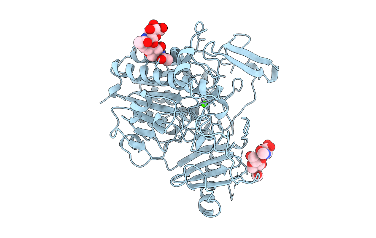
1FSU image
Deposition Date 1996-07-29
Release Date 1998-02-04
Last Version Date 2025-03-26
Entry Detail
PDB ID:
1FSU
Keywords:
Title:
Crystal Structure of 4-Sulfatase (human)
Biological Source:
Source Organism:
Homo sapiens (Taxon ID: 9606)
Host Organism:
Method Details:
Experimental Method:
Resolution:
2.50 Å
R-Value Free:
0.24
R-Value Work:
0.18
R-Value Observed:
0.18
Space Group:
P 41 21 2
Macromolecular Entities
Structures with similar UniProt ID
Protein Blast
Polymer Type:polypeptide(L)
Molecule:N-ACETYLGALACTOSAMINE-4-SULFATASE
Gene (Uniprot):ARSB
Chain IDs:A
Chain Length:492
Number of Molecules:1
Biological Source:Homo sapiens
Modified Residue
Compound ID Chain ID Parent Comp ID Details 2D Image
ALS A ALA (3S)-3-(SULFOOXY)-L-SERINE
ASN A ASN GLYCOSYLATION SITE
Primary Citation
Structure of a human lysosomal sulfatase.
Structure 5 277 289 (1997)
PMID: 9032078 DOI: 10.1016/S0969-2126(97)00185-8

Abstact

BACKGROUND . Sulfatases catalyze the hydrolysis of sulfuric acid esters from a wide variety of substrates including glycosaminoglycans, glycolipids and steroids. There is sufficient common sequence similarity within the class of sulfatase enzymes to indicate that they have a common structure. Deficiencies of specific lysosomal sulfatases that are involved in the degradation of glycosamino-glycans lead to rare inherited clinical disorders termed mucopolysaccharidoses. In sufferers of multiple sulfatase deficiency, all sulfatases are inactive because an essential post-translational modification of a specific active-site cysteine residue to oxo-alanine does not occur. Studies of this disorder have contributed to location and characterization of the sulfatase active site. To understand the catalytic mechanism of sulfatases, and ultimately the determinants of their substrate specificities, we have determined the structure of N-acetylgalactosamine-4-sulfatase. RESULTS . The crystal structure of the enzyme has been solved and refined at 2.5 resolution using data recorded at both 123K and 273K. The structure has two domains, the larger of which belongs to the alpha/beta class of proteins and contains the active site. The enzyme active site in the crystals contains several hitherto undescribed features. The active-site cysteine residue, Cys91, is found as the sulfate derivative of the aldehyde species, oxo-alanine. The sulfate is bound to a previously undetected metal ion, which we have identified as calcium. The structure of a vanadate-inhibited form of the enzyme has also been solved, and this structure shows that vanadate has replaced sulfate in the active site and that the vanadate is covalently linked to the protein. Preliminary data is presented for crystals soaked in the monosaccharide N-acetylgalactosamine, the structure of which forms a product complex of the enzyme. CONCLUSIONS . The structure of N-acetylgalactosamine-4-sulfatase reveals that residues conserved amongst the sulfatase family are involved in stabilizing the calcium ion and the sulfate ester in the active site. This suggests an archetypal fold for the family of sulfatases. A catalytic role is proposed for the post-translationally modified highly conserved cysteine residue. Despite a lack of any previously detectable sequence similarity to any protein of known structure, the large sulfatase domain that contains the active site closely resembles that of alkaline phosphatase: the calcium ion in sulfatase superposes on one of the zinc ions in alkaline phosphatase and the sulfate ester of Cys91 superposes on the phosphate ion found in the active site of alkaline phosphatase.

Legend

Protein

Chemical

Disease

Primary Citation of related structures