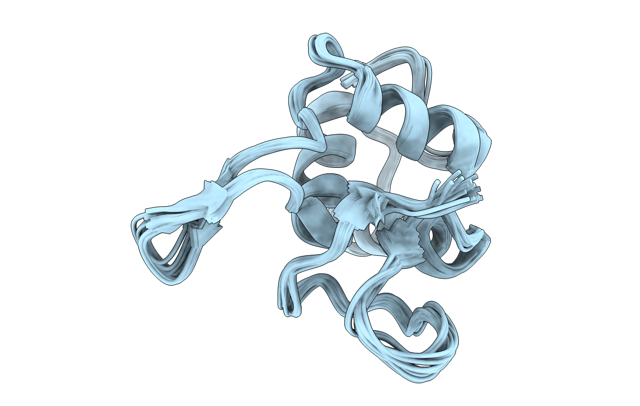
1FSH image
Deposition Date 2000-09-08
Release Date 2001-03-08
Last Version Date 2024-05-29
Entry Detail
PDB ID:
1FSH
Title:
STRUCTURAL BASIS OF THE RECOGNITION OF THE DISHEVELLED DEP DOMAIN IN THE WNT SIGNALING PATHWAY
Biological Source:
Source Organism:
Mus musculus (Taxon ID: 10090)
Host Organism:
Method Details:
Experimental Method:
Conformers Calculated:
1000
Conformers Submitted:
20
Selection Criteria:
target function
Macromolecular Entities
Polymer Type:polypeptide(L)
Molecule:DISHEVELLED-1
Gene (Uniprot):Dvl1
Chain IDs:A
Chain Length:105
Number of Molecules:1
Biological Source:Mus musculus
Ligand Molecules
Primary Citation
Structural basis of the recognition of the dishevelled DEP domain in the Wnt signaling pathway.
Nat.Struct.Biol. 7 1178 1184 (2000)
PMID: 11101902 DOI: 10.1038/82047

Abstact

The DEP domain of Dishevelled (Dvl) proteins transduces signals to effector proteins downstream of Dvl in the Wnt pathway. Here we report that DEP-containing mutants inhibit Wnt-induced, but not Dvl-induced, activation of the transcription factor Lef-1. This inhibitory effect is weakened by a K434M mutation. Nuclear magnetic resonance spectroscopy revealed that the DEP domain of mouse Dvl1 comprises a three-helix bundle, a beta-hairpin 'arm' and two short beta-strands at the C-terminal region. Lys 434 is located at the tip of the beta-hairpin 'arm'. Based on our findings, we conclude that DEP interacts with regulators upstream of Dvl via a strong electric dipole on the molecule's surface created by Lys 434, Asp 445 and Asp 448; the electric dipole and the putative membrane binding site are at two different locations.

Legend

Protein

Chemical

Disease

Primary Citation of related structures