1FRP image
Deposition Date 1994-08-26
Release Date 1994-11-30
Last Version Date 2024-02-07
Entry Detail
PDB ID:
1FRP
Title:
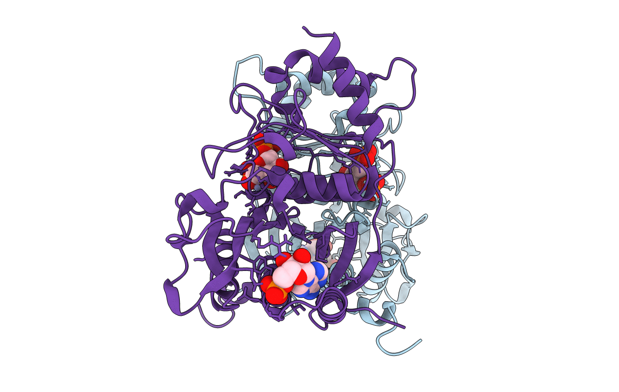
CRYSTAL STRUCTURE OF FRUCTOSE-1,6-BISPHOSPHATASE COMPLEXED WITH FRUCTOSE-2,6-BISPHOSPHATE, AMP AND ZN2+ AT 2.0 ANGSTROMS RESOLUTION. ASPECTS OF SYNERGISM BETWEEN INHIBITORS
Biological Source:
Source Organism:
Sus scrofa (Taxon ID: 9823)
Method Details:
Experimental Method:
Resolution:
2.00 Å
R-Value Work:
0.18
R-Value Observed:
0.18
Space Group:
P 21 21 2
Macromolecular Entities
Polymer Type:polypeptide(L)
Molecule:FRUCTOSE 1,6-BISPHOSPHATASE
Gene (Uniprot):FBP1
Chain IDs:A, B
Chain Length:335
Number of Molecules:2
Biological Source:Sus scrofa
Primary Citation
Crystal structure of fructose-1,6-bisphosphatase complexed with fructose 2,6-bisphosphate, AMP, and Zn2+ at 2.0-A resolution: aspects of synergism between inhibitors.
Proc.Natl.Acad.Sci.USA 91 12482 12486 (1994)
PMID: 7809062 DOI: 10.1073/pnas.91.26.12482

Abstact

The crystal structure of fructose-1,6-bisphosphatase (Fru-1,6-Pase; EC 3.1.3.11) complexed with Zn2+ and two allosteric regulators, AMP and fructose 2,6-bisphosphate (Fru-2,6-P2) has been determined at 2.0-A resolution. In the refined model, the crystallographic R factor is 0.189 with rms deviations of 0.014 A and 2.8 degrees from ideal geometries for bond lengths and bond angles, respectively. A 15 degrees rotation is observed between the upper dimer C1C2 and the lower dimer C3C4 relative to the R-form structure (fructose 6-phosphate complex), consistent with that expected from a T-form structure. The major difference between the structure of the previously determined Fru-2,6-P2 complex (R form) and that of the current quaternary T-form complex lies in the active site domain. A zinc binding site distinct from the three binding sites established earlier was identified within each monomer. Helix H4 (residues 123-127) was found to be better defined than in previously studied ligated Fru-1,6-Pase structures. Interactions between monomers in the active site domain were found involving H4 residues from one monomer and residues Tyr-258 and Arg-243 from the adjacent monomer. Cooperativity between AMP and Fru-2,6-P2 in signal transmission probably involves the following features: an AMP site, the adjacent B3 strand (residues 113-118), the metal site, the immediate active site, the short helix H4 (residues 123-127), and Tyr-258 and Arg-243 from the adjacent monomer within the upper (or lower) dimer. The closest distance between the immediate active site and that on the adjacent monomer is only 5 A. Thus, the involvement of H4 in signal transmission adds another important pathway to the scheme of the allosteric mechanism of Fru-1,6-Pase.

Legend

Protein

Chemical

Disease

Primary Citation of related structures