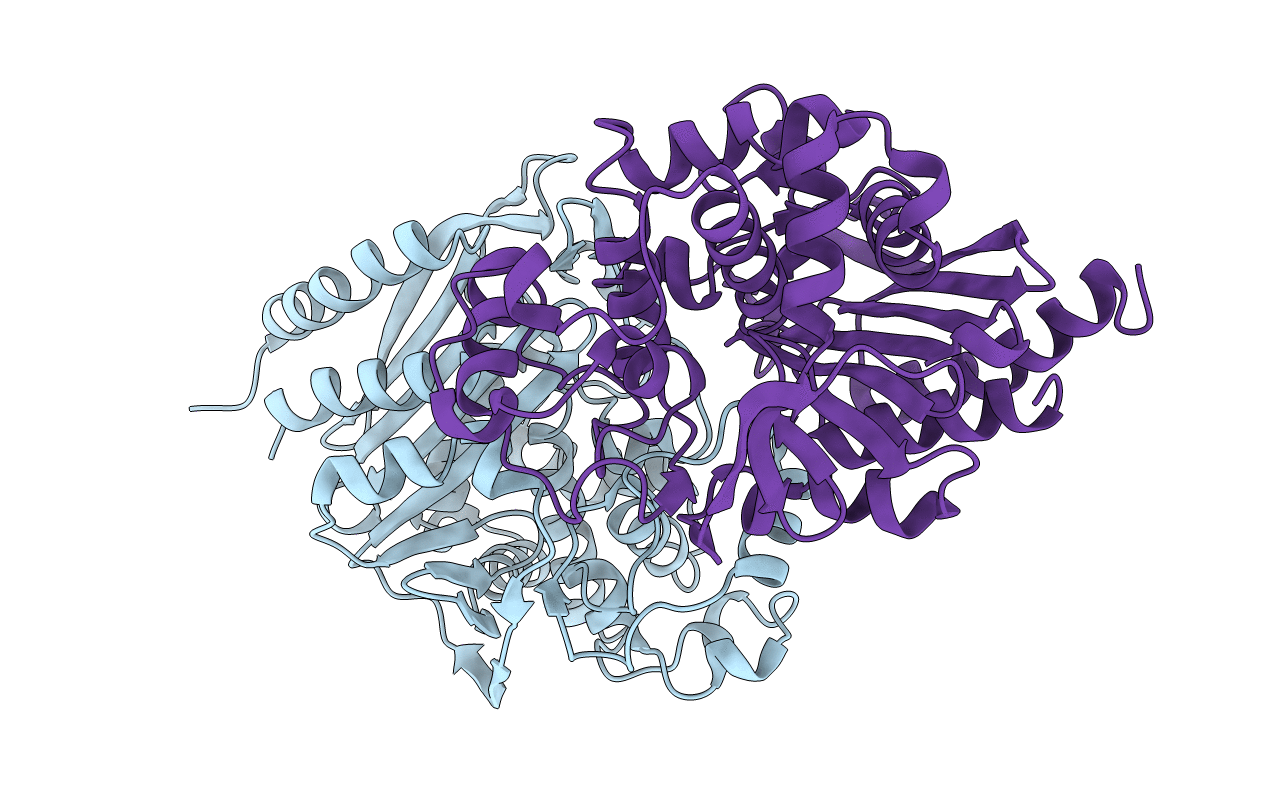
1FR1 image
Deposition Date 2000-09-07
Release Date 2001-01-17
Last Version Date 2024-02-07
Entry Detail
PDB ID:
1FR1
Keywords:
Title:
REFINED CRYSTAL STRUCTURE OF BETA-LACTAMASE FROM CITROBACTER FREUNDII INDICATES A MECHANISM FOR BETA-LACTAM HYDROLYSIS
Biological Source:
Source Organism:
Method Details:
Experimental Method:
Resolution:
2.00 Å
R-Value Observed:
0.18
Space Group:
P 21 21 21
Macromolecular Entities
Polymer Type:polypeptide(L)
Molecule:BETA-LACTAMASE
Chain IDs:A, B
Chain Length:361
Number of Molecules:2
Biological Source:Citrobacter freundii
Primary Citation
Refined crystal structure of beta-lactamase from Citrobacter freundii indicates a mechanism for beta-lactam hydrolysis.
Nature 343 284 288 (1990)
PMID: 2300174 DOI: 10.1038/343284a0

Abstact

Beta-Lactamases (EC 3.5.2.6, 'penicillinases') are a family of enzymes that protect bacteria against the lethal effects of cell-wall synthesis of penicillins, cephalosporins and related antibiotic agents, by hydrolysing the beta-lactam antibiotics to biologically inactive compounds. Their production can, therefore, greatly contribute to the clinical problem of antibiotic resistance. Three classes of beta-lactamases--A, B and C--have been identified on the basis of their amino-acid sequence; class B beta-lactamases are metalloenzymes, and are clearly distinct from members of class A and C beta-lactamases, which both contain an active-site serine residue involved in the formation of an acyl enzyme with beta-lactam substrates during catalysis. It has been predicted that class C beta-lactamases share common structural features with D,D-carboxypeptidases and class A beta-lactamases, and further, suggested that class A and class C beta-lactamases have the same evolutionary origin as other beta-lactam target enzymes. We report here the refined three-dimensional structure of the class C beta-lactamase from Citrobacter freundii at 2.0-A resolution and confirm the predicted structural similarity. The refined structure of the acyl-enzyme formed with the monobactam inhibitor aztreonam at 2.5-A resolution defines the enzyme's active site and, along with molecular modelling, indicates a mechanism for beta-lactam hydrolysis. This leads to the hypothesis that Tyr 150 functions as a general base during catalysis.

Legend

Protein

Chemical

Disease

Primary Citation of related structures