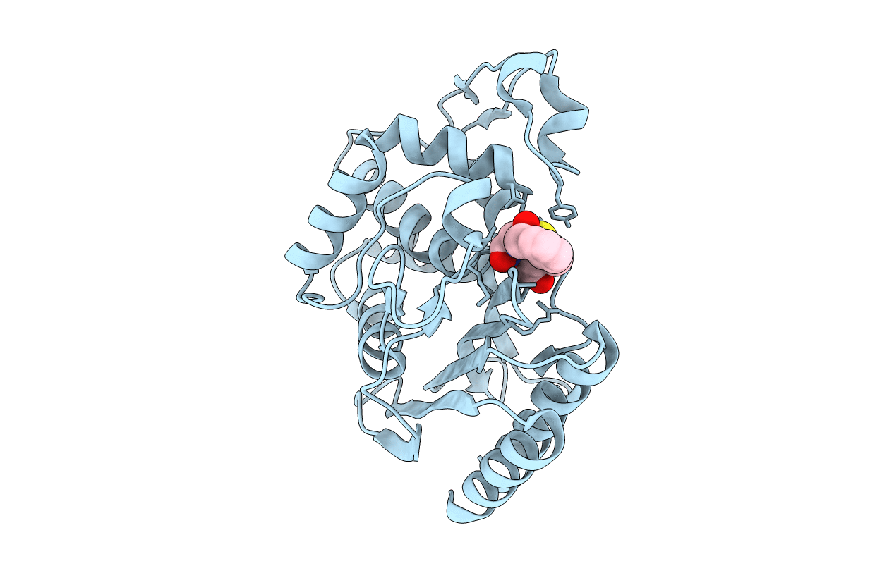
1FQG image
Deposition Date 2000-09-05
Release Date 2000-11-01
Last Version Date 2024-10-30
Entry Detail
PDB ID:
1FQG
Keywords:
Title:
MOLECULAR STRUCTURE OF THE ACYL-ENZYME INTERMEDIATE IN TEM-1 BETA-LACTAMASE
Biological Source:
Source Organism:
Escherichia coli (Taxon ID: 562)
Host Organism:
Method Details:
Experimental Method:
Resolution:
1.70 Å
R-Value Work:
0.18
R-Value Observed:
0.18
Space Group:
P 21 21 21
Macromolecular Entities
Polymer Type:polypeptide(L)
Molecule:TEM-1 BETA-LACTAMASE
Gene (Uniprot):bla, blaT-3, blaT-4, blaT-5, blaT-6
Mutations:GLU166ASN MUTATION
Chain IDs:A
Chain Length:263
Number of Molecules:1
Biological Source:Escherichia coli
Ligand Molecules
Primary Citation
Molecular structure of the acyl-enzyme intermediate in beta-lactam hydrolysis at 1.7 A resolution.
Nature 359 700 705 (1992)
PMID: 1436034 DOI: 10.1038/359700a0

Abstact

The X-ray crystal structure of the molecular complex of penicillin G with a deacylation-defective mutant of the RTEM-1 beta-lactamase from Escherichia coli shows how these antibiotics are recognized and destroyed. Penicillin G is covalently bound to Ser 70 0 gamma as an acyl-enzyme intermediate. The deduced catalytic mechanism uses Ser 70 0 gamma as the attacking nucleophile during acylation. Lys 73 N zeta acts as a general base in abstracting a proton from Ser 70 and transferring it to the thiazolidine ring nitrogen atom via Ser 130 0 gamma. Deacylation is accomplished by nucleophilic attack on the penicilloyl carbonyl carbon by a water molecule assisted by the general base, Glu 166.

Legend

Protein

Chemical

Disease

Primary Citation of related structures