Deposition Date
2000-08-31
Release Date
2001-03-07
Last Version Date
2024-10-16
Entry Detail
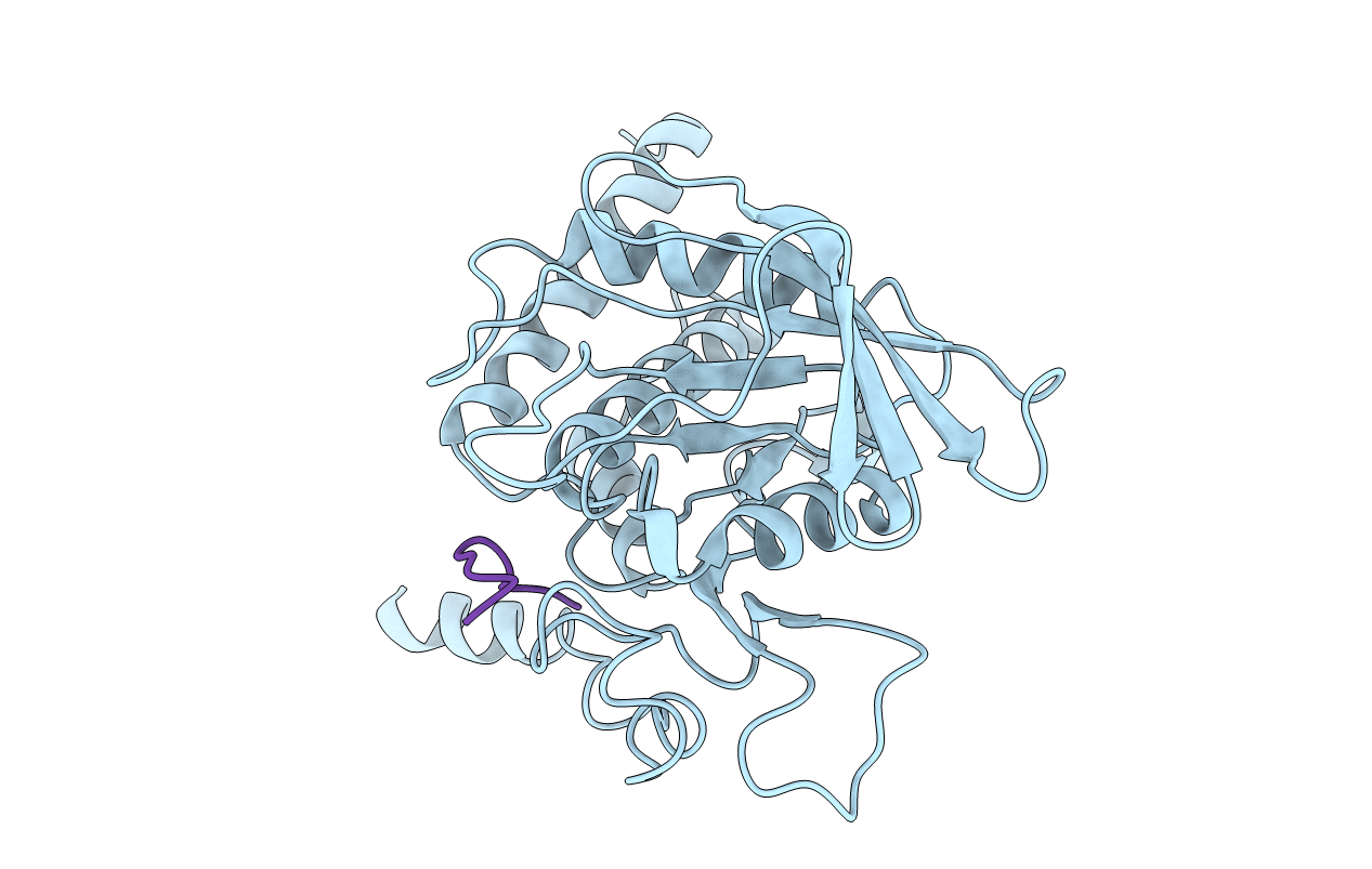
PDB ID:
1FPR
Keywords:
Title:
CRYSTAL STRUCTURE OF THE COMPLEX FORMED BETWEEN THE CATALYTIC DOMAIN OF SHP-1 AND AN IN VITRO PEPTIDE SUBSTRATE PY469 DERIVED FROM SHPS-1.
Biological Source:
Source Organism(s):
Homo sapiens (Taxon ID: 9606)
Expression System(s):
Method Details:
Experimental Method:
Resolution:
2.50 Å
R-Value Free:
0.30
R-Value Work:
0.19
R-Value Observed:
0.19
Space Group:
P 21 21 2