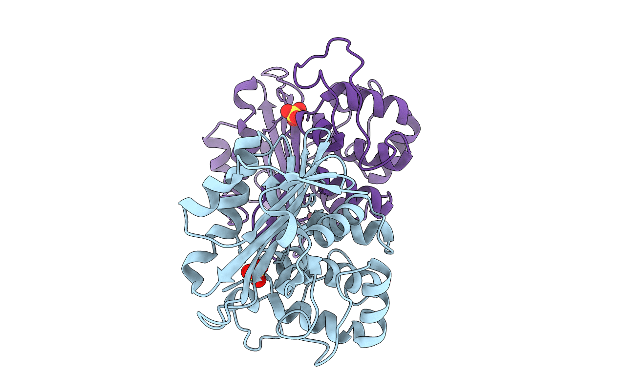
1FOF image
Entry Detail
PDB ID:
1FOF
Keywords:
Title:
CRYSTAL STRUCTURE OF THE CLASS D BETA-LACTAMASE OXA-10
Biological Source:
Source Organism:
Host Organism:
PDB Version:
Deposition Date:
2000-08-28
Release Date:
2000-10-09
Method Details:
Experimental Method:
Resolution:
2.00 Å
R-Value Free:
0.20
R-Value Work:
0.17
R-Value Observed:
0.17
Space Group:
P 21 21 21
Macromolecular Entities
Polymer Type:polypeptide(L)
Description:BETA LACTAMASE OXA-10
Chain IDs:A, B
Chain Length:246
Number of Molecules:2
Biological Source:Pseudomonas aeruginosa
Primary Citation
Crystal structure of the class D beta-lactamase OXA-10.
Nat.Struct.Biol. 7 918 925 (2000)
PMID: 11017203 DOI: 10.1038/79688

Abstact

We report the crystal structure of a class D beta-lactamase, the broad spectrum enzyme OXA-10 from Pseudomonas aeruginosa at 2.0 A resolution. There are significant differences between the overall fold observed in this structure and those of the evolutionarily related class A and class C beta-lactamases. Furthermore, the structure suggests the unique, cation mediated formation of a homodimer. Kinetic and hydrodynamic data shows that the dimer is a relevant species in solution and is the more active form of the enzyme. Comparison of the molecular details of the active sites of the class A and class C enzymes with the OXA-10 structure reveals that there is no counterpart in OXA-10 to the residues proposed to act as general bases in either of these enzymes (Glu 166 and Tyr 150, respectively). Our structures of the native and chloride inhibited forms of OXA-10 suggest that the class D enzymes have evolved a distinct catalytic mechanism for beta-lactam hydrolysis. Clinical variants of OXA-10 are also discussed in light of the structure.

Legend

Protein

Chemical

Disease

Primary Citation of related structures