Deposition Date
2000-08-17
Release Date
2000-11-01
Last Version Date
2024-11-06
Entry Detail
PDB ID:
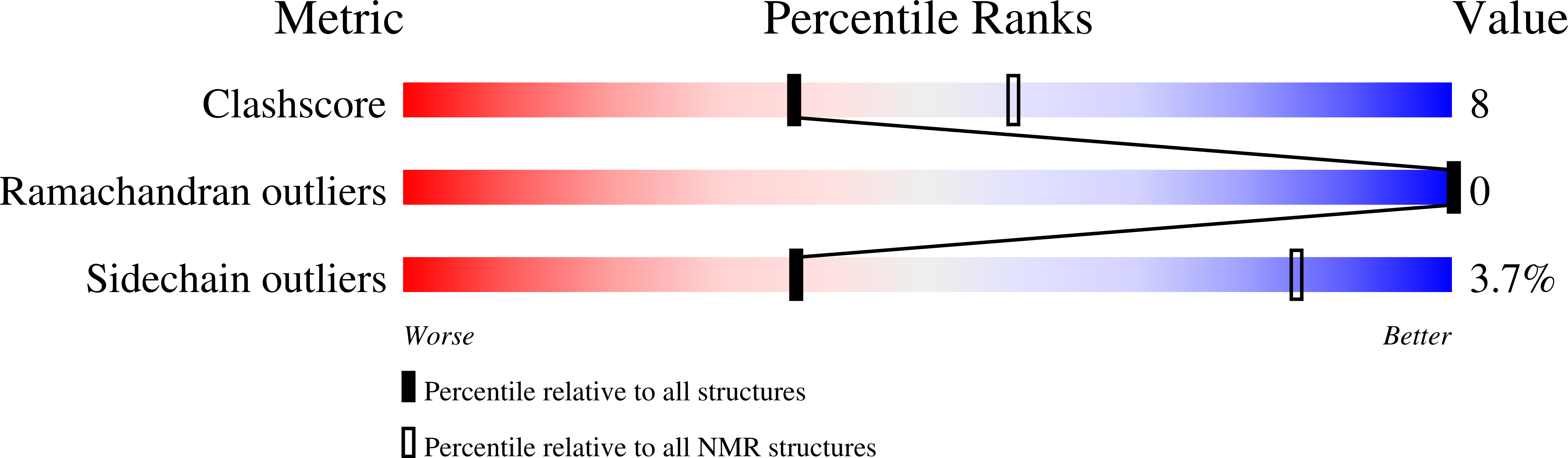
1FMH
Keywords:
Title:
NMR SOLUTION STRUCTURE OF A DESIGNED HETERODIMERIC LEUCINE ZIPPER
Method Details:
Experimental Method:
Conformers Calculated:
50
Conformers Submitted:
25
Selection Criteria:
structures with the least restraint violations,structures with the lowest energy