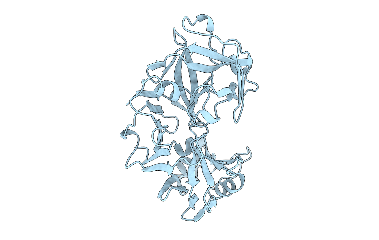
1FLH image
Deposition Date 2000-08-14
Release Date 2001-10-31
Last Version Date 2024-10-09
Entry Detail
PDB ID:
1FLH
Keywords:
Title:
CRYSTAL STRUCTURE OF HUMAN UROPEPSIN AT 2.45 A RESOLUTION
Biological Source:
Source Organism:
Homo sapiens (Taxon ID: 9606)
Method Details:
Experimental Method:
Resolution:
2.45 Å
R-Value Work:
0.16
R-Value Observed:
0.16
Space Group:
P 21 21 21
Macromolecular Entities
Structures with similar UniProt ID
Protein Blast
Polymer Type:polypeptide(L)
Molecule:UROPEPSIN
Chain IDs:A
Chain Length:326
Number of Molecules:1
Biological Source:Homo sapiens
Primary Citation
Structure of human uropepsin at 2.45 A resolution.
Acta Crystallogr.,Sect.D 57 1560 1570 (2001)
PMID: 11679720 DOI: 10.1107/S0907444901013865

Abstact

The molecular structure of human uropepsin, an aspartic proteinase from the urine produced in the form of pepsinogen A in the gastric mucosa, has been determined by molecular replacement using human pepsin as the search model. Crystals belong to space group P2(1)2(1)2(1), with unit-cell parameters a = 50.99, b = 75.56, c = 89.90 A. Crystallographic refinement led to an R factor of 0.161 at 2.45 A resolution. The positions of 2437 non-H protein atoms in 326 residues have been determined and the model contains 143 water molecules. The structure is bilobal, consisting of two predominantly beta-sheet lobes which, as observed in other aspartic proteinases, are related by a pseudo-twofold axis. A model of the uropepsin-pepstatin complex has been constructed based on the high-resolution crystal structure of pepsin complexed with pepstatin.

Legend

Protein

Chemical

Disease

Primary Citation of related structures