Deposition Date
1999-02-22
Release Date
2000-03-01
Last Version Date
2024-11-06
Entry Detail
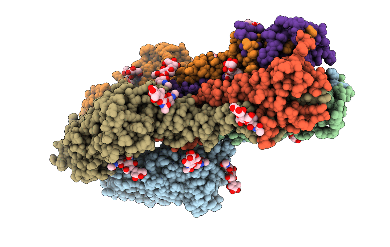
PDB ID:
1FLC
Keywords:
Title:
X-RAY STRUCTURE OF THE HAEMAGGLUTININ-ESTERASE-FUSION GLYCOPROTEIN OF INFLUENZA C VIRUS
Biological Source:
Source Organism(s):
Influenza C virus (C/Johannesburg/1/66) (Taxon ID: 100673)
Method Details:
Experimental Method:
Resolution:
3.20 Å
R-Value Free:
0.26
R-Value Work:
0.22
R-Value Observed:
0.22
Space Group:
P 43 2 2