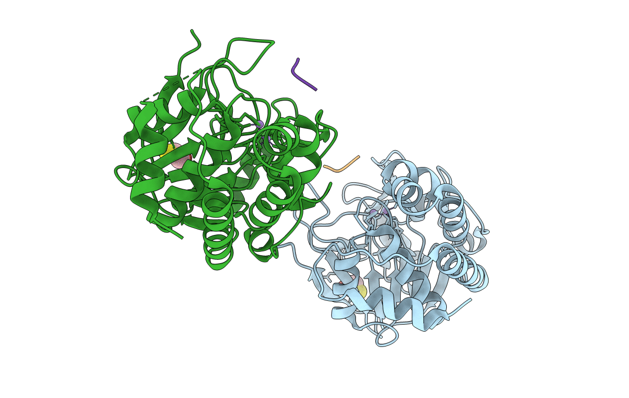
1FJM image
Entry Detail
PDB ID:
1FJM
Title:
Protein serine/threonine phosphatase-1 (alpha isoform, type 1) complexed with microcystin-LR toxin
Biological Source:
PDB Version:
Deposition Date:
1995-12-17
Release Date:
1996-06-20
Method Details:
Experimental Method:
Resolution:
2.10 Å
R-Value Work:
0.17
R-Value Observed:
0.17
Space Group:
P 21 21 21
Macromolecular Entities
Polymer Type:polypeptide(L)
Description:PROTEIN SERINE/THREONINE PHOSPHATASE-1 (ALPHA ISOFORM, TYPE 1)
Chain IDs:A, B
Chain Length:330
Number of Molecules:2
Biological Source:Oryctolagus cuniculus
Polymer Type:polypeptide(L)
Description:microcystin LR
Chain IDs:C (auth: M), D (auth: N)
Chain Length:7
Number of Molecules:2
Biological Source:Microcystis aeruginosa
Peptide-like Molecules
PRD_000212
Primary Citation
Three-dimensional structure of the catalytic subunit of protein serine/threonine phosphatase-1.
Nature 376 745 753 (1995)
PMID: 7651533 DOI: 10.1038/376745a0

Abstact

The crystal structure of mammalian protein phosphatase-1, complexed with the toxin microcystin and determined at 2.1 A resolution, reveals that it is a metalloenzyme unrelated in architecture to the tyrosine phosphatases. Two metal ions are positioned by a central beta-alpha-beta-alpha-beta scaffold at the active site, from which emanate three surface grooves that are potential binding sites for substrates and inhibitors. The carboxy terminus is positioned at the end of one of the grooves such that regulatory sequences following the domain might modulate function. The fold of the catalytic domain is expected to be closely preserved in protein phosphatases 2A and 2B (calcineurin).

Legend

Protein

Chemical

Disease

Primary Citation of related structures