
Deposition Date
2000-08-08
Release Date
2001-07-18
Last Version Date
2024-10-16
Entry Detail
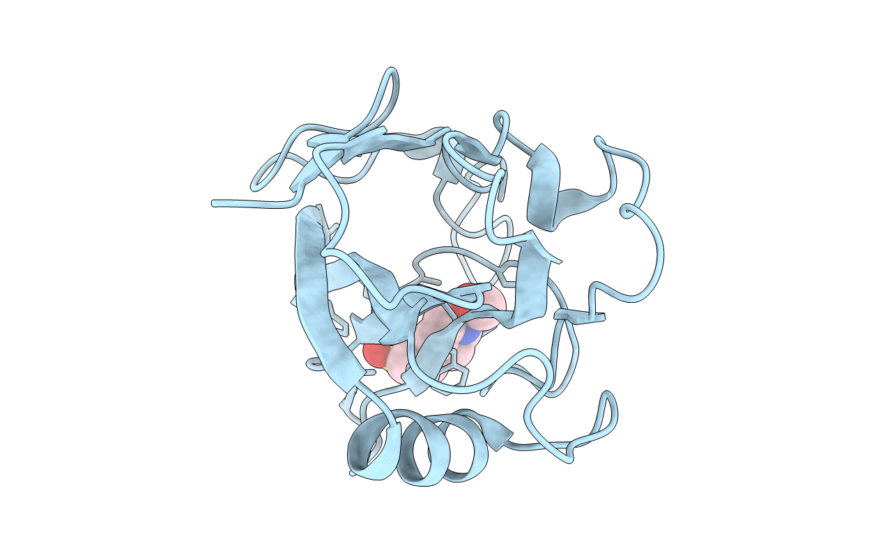
PDB ID:
1FJJ
Keywords:
Title:
CRYSTAL STRUCTURE OF E.COLI YBHB PROTEIN, A NEW MEMBER OF THE MAMMALIAN PEBP FAMILY
Biological Source:
Source Organism(s):
Escherichia coli (Taxon ID: 562)
Method Details:
Experimental Method:
Resolution:
1.66 Å
R-Value Free:
0.19
R-Value Work:
0.17
Space Group:
P 32 2 1
