Deposition Date
2000-08-08
Release Date
2001-08-08
Last Version Date
2024-05-22
Entry Detail
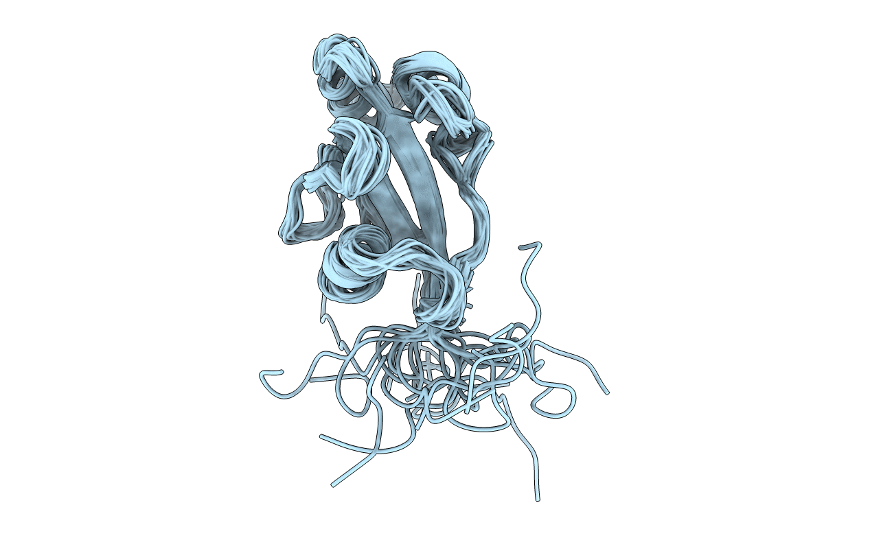
PDB ID:
1FJD
Keywords:
Title:
HUMAN PARVULIN-LIKE PEPTIDYL PROLYL CIS/TRANS ISOMERASE, HPAR14
Biological Source:
Source Organism(s):
Homo sapiens (Taxon ID: 9606)
Expression System(s):
Method Details:
Experimental Method:
Conformers Calculated:
100
Conformers Submitted:
20
Selection Criteria:
structures with the lowest energy