1FIR image
Deposition Date 2000-08-06
Release Date 2001-01-17
Last Version Date 2023-08-02
Entry Detail
PDB ID:
1FIR
Keywords:
Title:
CRYSTAL STRUCTURE OF HIV-1 REVERSE TRANSCRIPTION PRIMER TRNA(LYS3)
Biological Source:
Source Organism:
Bos taurus (Taxon ID: 9913)
Method Details:
Experimental Method:
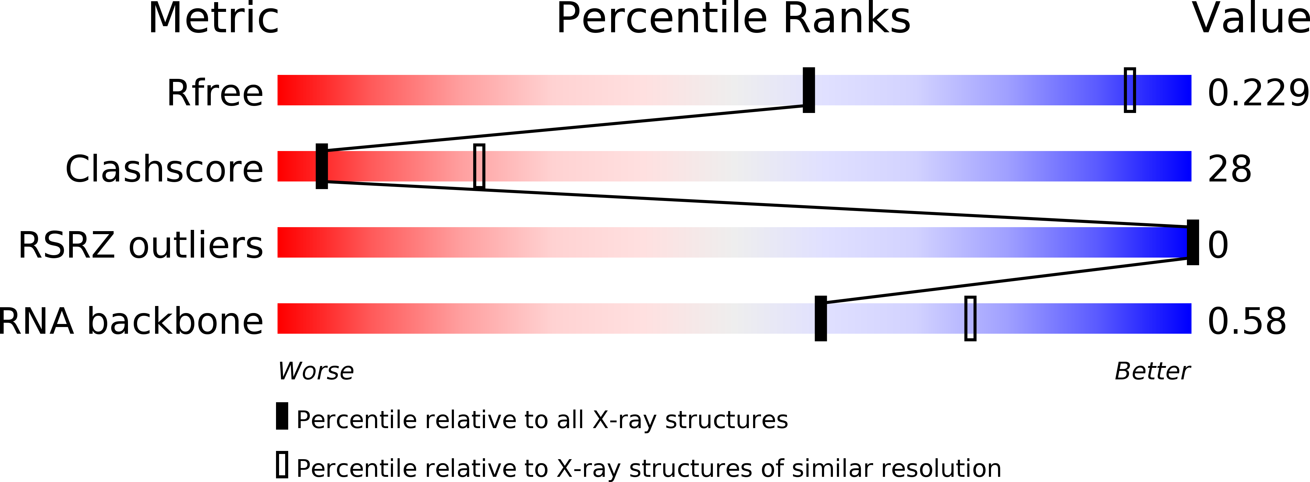
Resolution:
3.30 Å
R-Value Free:
0.22
R-Value Work:
0.18
R-Value Observed:
0.18
Space Group:
P 32 1 2
Macromolecular Entities
Polymer Type:polyribonucleotide
Molecule:HIV-1 REVERSE TRANSCRIPTION PRIMER TRNA(LYS3)
Chain IDs:A
Chain Length:76
Number of Molecules:1
Biological Source:Bos taurus
Modified Residue
Compound ID Chain ID Parent Comp ID Details 2D Image
12A A A ?
1MA A A ?
2MG A G 2N-METHYLGUANOSINE-5'-MONOPHOSPHATE
2MU A U 2',5-DIMETHYLURIDINE-5'-MONOPHOSPHATE
5MC A C 5-METHYLCYTIDINE-5'-MONOPHOSPHATE
70U A U ?
7MG A G ?
H2U A U 5,6-DIHYDROURIDINE-5'-MONOPHOSPHATE
PSU A U PSEUDOURIDINE-5'-MONOPHOSPHATE
Primary Citation
The crystal structure of HIV reverse-transcription primer tRNA(Lys,3) shows a canonical anticodon loop.
RNA 6 1347 1355 (2000)
PMID: 11073212 DOI: 10.1017/S1355838200000911

Abstact

We have solved to 3.3 A resolution the crystal structure of the HIV reverse-transcription primer tRNA(Lys,3). The overall structure is exactly comparable to the well-known L-shape structure first revealed by yeast tRNA(Phe). In particular, it unambiguously shows a canonical anticodon loop. This contradicts previous results in short RNA fragment studies and leads us to conclude that neither frameshifting specificities of tRNA(Lys) nor tRNA(Lys,3) primer selection by HIV are due to a specific three-dimensional anticodon structure. Comparison of our structure with the results of an NMR study on a hairpin representing a nonmodified anticodon stem-loop makes plausible the conclusion that chemical modifications of the wobble base U34 to 5-methoxycarbonyl-methyl-2-thiouridine and of A37 to 2-methylthio-N-6-threonylcarbamoyl-adenosine would be responsible for a canonical 7-nt anticodon-loop structure, whereas the unmodified form would result in a noncanonical UUU short triloop. The hexagonal crystal packing is remarkable and shows tight dimers of tRNAs forming a right-handed double superhelix. Within the dimers, the tRNAs are associated head-to-tail such that the CCA end of one tRNA interacts with the anticodon of the symmetry-related tRNA. This provides us with a partial view of a codon-anticodon interaction and gives insights into the positioning of residue 37, and of its posttranscriptional modifications, relative to the first base of the codon.

Legend

Protein

Chemical

Disease

Primary Citation of related structures