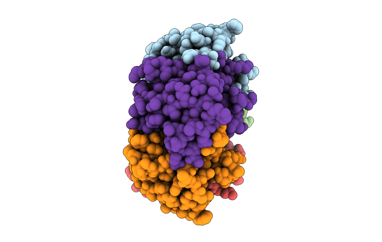
Cholera toxin, a heterohexameric AB5 enterotoxin released by Vibrio cholera, induces a profuse secretory diarrhea in susceptible hosts. Choleragenoid, the B subunit pentamer of cholera toxin, directs the enzymatic A subunit to its target by binding the GM1 gangliosides exposed on the luminal surface of intestinal epithelial cells. The crystal structure of choleragenoid has been independently solved and refined at 2.4 A resolution by combining single isomorphous replacement with non-crystallographic symmetry averaging. The structure of the B subunits, and their pentameric arrangement, closely resembles that reported for the intact holotoxin, choleragen, the heat-labile enterotoxin from Escherichia coli, and for a choleragenoid-GM1 pentasaccharide complex. In the absence of the A subunit the central cavity of the B pentamer is a highly solvated channel. The binding of choleragenoid to the A subunit or to its receptor pentasaccharide modestly affects the local stereochemistry without perceptibly altering the subunit interface.
Legend
Protein
Chemical
Disease