1FE1 image
Deposition Date 2000-07-20
Release Date 2001-02-14
Last Version Date 2024-12-25
Entry Detail
PDB ID:
1FE1
Title:
CRYSTAL STRUCTURE PHOTOSYSTEM II
Biological Source:
Source Organism:
Method Details:
Experimental Method:
Resolution:
3.80 Å
Space Group:
P 21 21 21
Macromolecular Entities
Polymer Type:polypeptide(L)
Molecule:PROTEIN (PHOTOSYSTEM II: SUBUNIT PSBA)
Chain IDs:A, J
Chain Length:169
Number of Molecules:2
Biological Source:Synechococcus elongatus
Polymer Type:polypeptide(L)
Molecule:PROTEIN (PHOTOSYSTEM II: SUBUNIT PSBD)
Chain IDs:B, K
Chain Length:174
Number of Molecules:2
Biological Source:Synechococcus elongatus
Polymer Type:polypeptide(L)
Molecule:PROTEIN (PHOTOSYSTEM II: SUBUNIT PSBC)
Chain IDs:C, L
Chain Length:156
Number of Molecules:2
Biological Source:Synechococcus elongatus
Polymer Type:polypeptide(L)
Molecule:PROTEIN (PHOTOSYSTEM II: SUBUNIT PSBB)
Chain IDs:D, M
Chain Length:155
Number of Molecules:2
Biological Source:Synechococcus elongatus
Polymer Type:polypeptide(L)
Molecule:PROTEIN (PHOTOSYSTEM II: SUBUNIT PSBE)
Chain IDs:E, N
Chain Length:40
Number of Molecules:2
Biological Source:Synechococcus elongatus
Polymer Type:polypeptide(L)
Molecule:PROTEIN (PHOTOSYSTEM II: SUBUNIT PSBF)
Chain IDs:F, O
Chain Length:30
Number of Molecules:2
Biological Source:Synechococcus elongatus
Polymer Type:polypeptide(L)
Molecule:PROTEIN (PHOTOSYSTEM II: SUBUNIT UNKNOWN)
Chain IDs:G, P
Chain Length:312
Number of Molecules:2
Biological Source:Synechococcus elongatus
Polymer Type:polypeptide(L)
Molecule:PROTEIN (PHOTOSYSTEM II: SUBUNIT PSBO)
Chain IDs:H, Q
Chain Length:115
Number of Molecules:2
Biological Source:Synechococcus elongatus
Polymer Type:polypeptide(L)
Molecule:PROTEIN (PHOTOSYSTEM II: SUBUNIT PSBV)
Chain IDs:I, R
Chain Length:87
Number of Molecules:2
Biological Source:Synechococcus elongatus
Primary Citation
Crystal structure of photosystem II from Synechococcus elongatus at 3.8 A resolution.
Nature 409 739 743 (2001)
PMID: 11217865 DOI: 10.1038/35055589

Abstact

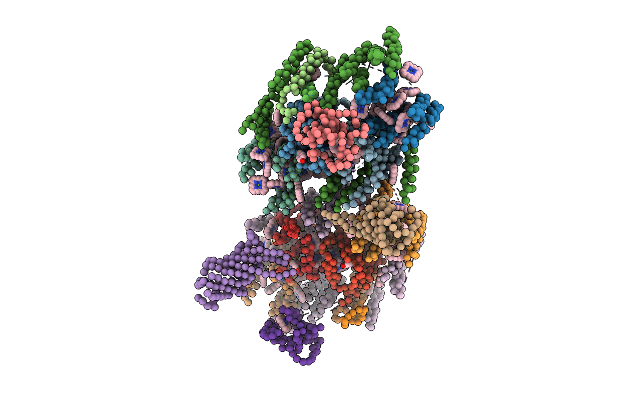
Oxygenic photosynthesis is the principal energy converter on earth. It is driven by photosystems I and II, two large protein-cofactor complexes located in the thylakoid membrane and acting in series. In photosystem II, water is oxidized; this event provides the overall process with the necessary electrons and protons, and the atmosphere with oxygen. To date, structural information on the architecture of the complex has been provided by electron microscopy of intact, active photosystem II at 15-30 A resolution, and by electron crystallography on two-dimensional crystals of D1-D2-CP47 photosystem II fragments without water oxidizing activity at 8 A resolution. Here we describe the X-ray structure of photosystem II on the basis of crystals fully active in water oxidation. The structure shows how protein subunits and cofactors are spatially organized. The larger subunits are assigned and the locations and orientations of the cofactors are defined. We also provide new information on the position, size and shape of the manganese cluster, which catalyzes water oxidation.

Legend

Protein

Chemical

Disease

Primary Citation of related structures