1FDP image
Deposition Date 1998-12-03
Release Date 1999-12-03
Last Version Date 2024-11-20
Entry Detail
PDB ID:
1FDP
Keywords:
Title:
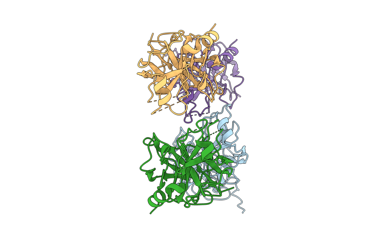
PROENZYME OF HUMAN COMPLEMENT FACTOR D, RECOMBINANT PROFACTOR D
Biological Source:
Source Organism:
Homo sapiens (Taxon ID: 9606)
Host Organism:
Method Details:
Experimental Method:
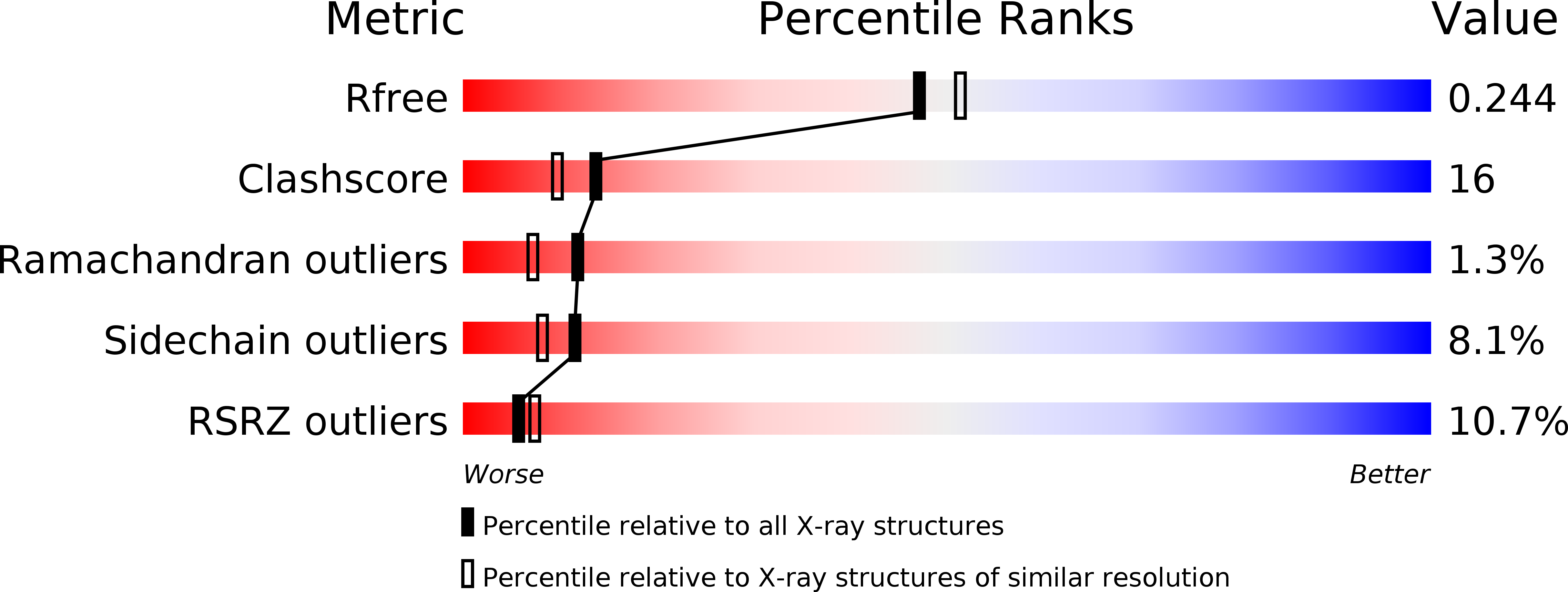
Resolution:
2.10 Å
R-Value Free:
0.25
R-Value Work:
0.20
R-Value Observed:
0.20
Space Group:
P 1 21 1
Macromolecular Entities
Polymer Type:polypeptide(L)
Molecule:PROENZYME OF COMPLEMENT FACTOR D
Gene (Uniprot):CFD
Chain IDs:A, B, C, D
Chain Length:235
Number of Molecules:4
Biological Source:Homo sapiens
Primary Citation

Abstact

The crystal structure of profactor D, determined at 2.1 A resolution with an Rfree and an R-factor of 25.1 and 20.4%, respectively, displays highly flexible or disordered conformation for five regions: N-22, 71-76, 143-152, 187-193 and 215-223. A comparison with the structure of its mature serine protease, complement factor D, revealed major conformational changes in the similar regions. Comparisons with the zymogen-active enzyme pairs of chymotrypsinogen, trypsinogen and prethrombin-2 showed a similar distribution of the flexible regions. However, profactor D is the most flexible of the four, and its mature enzyme displays inactive, self-inhibited active site conformation. Examination of the surface properties of the N-terminus-binding pocket indicates that Ile16 may play the initial positioning role for the N-terminus, and Leu17 probably also helps in inducing the required conformational changes. This process, perhaps shared by most chymotrypsinogen-like zymogens, is followed by a factor D-unique step, the re-orientation of an external Arg218 to an internal position for salt-bridging with Asp189, leading to the generation of the self-inhibited factor D.

Legend

Protein

Chemical

Disease

Primary Citation of related structures