Deposition Date
1993-04-20
Release Date
1993-10-31
Last Version Date
2024-02-07
Entry Detail
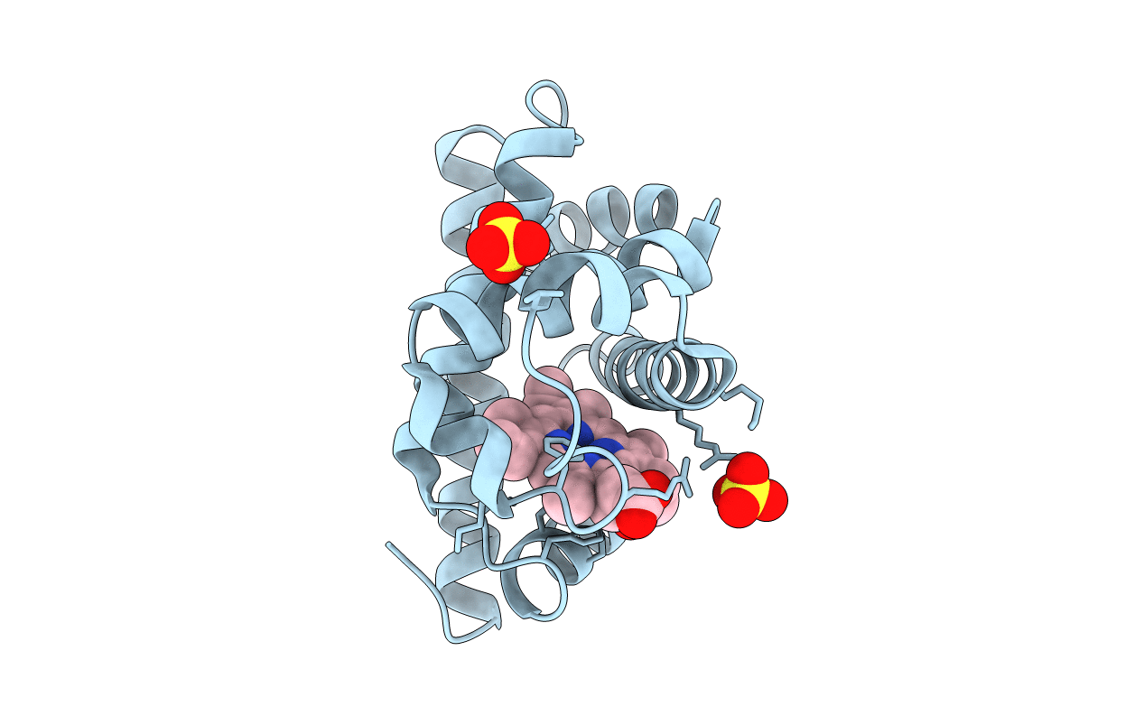
PDB ID:
1FCS
Keywords:
Title:
CRYSTAL STRUCTURE OF A DISTAL SITE DOUBLE MUTANT OF SPERM WHALE MYOGLOBIN AT 1.6 ANGSTROMS RESOLUTION
Biological Source:
Source Organism(s):
Physeter catodon (Taxon ID: 9755)
Method Details: