1FCH image
Deposition Date 2000-07-18
Release Date 2000-12-06
Last Version Date 2024-02-07
Entry Detail
PDB ID:
1FCH
Title:
CRYSTAL STRUCTURE OF THE PTS1 COMPLEXED TO THE TPR REGION OF HUMAN PEX5
Biological Source:
Source Organism:
Homo sapiens (Taxon ID: 9606)
(Taxon ID: )
Host Organism:
Method Details:
Experimental Method:
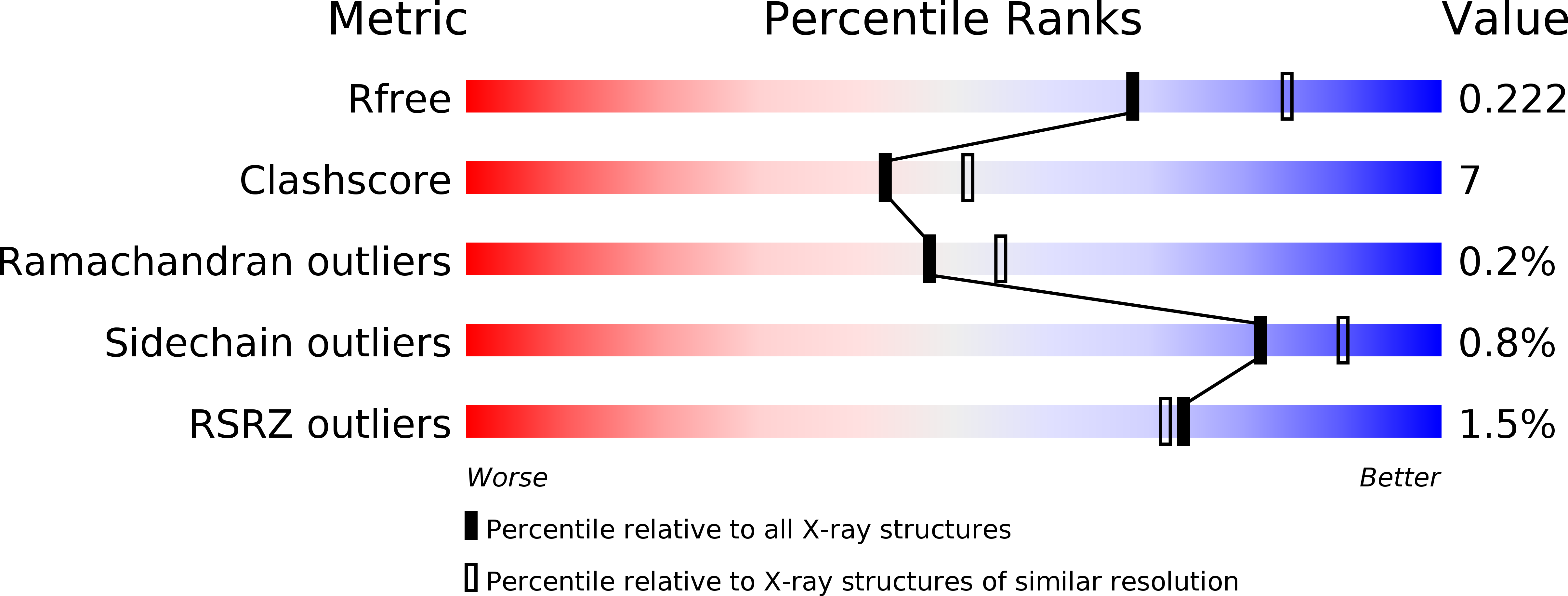
Resolution:
2.20 Å
R-Value Free:
0.22
R-Value Work:
0.19
R-Value Observed:
0.19
Space Group:
C 1 2 1
Macromolecular Entities
Polymer Type:polypeptide(L)
Molecule:PEROXISOMAL TARGETING SIGNAL 1 RECEPTOR
Gene (Uniprot):PEX5
Chain IDs:A, B
Chain Length:368
Number of Molecules:2
Biological Source:Homo sapiens
Polymer Type:polypeptide(L)
Molecule:PTS1-CONTAINING PEPTIDE
Chain IDs:C, D
Chain Length:5
Number of Molecules:2
Biological Source:
Primary Citation
Peroxisomal targeting signal-1 recognition by the TPR domains of human PEX5.
Nat.Struct.Biol. 7 1091 1095 (2000)
PMID: 11101887 DOI: 10.1038/81930

Abstact

Many proteins contain targeting signals within their sequences that specify their delivery to particular organelles. The peroxisomal targeting signal-1 (PTS1) is a C-terminal tripeptide that is sufficient to direct proteins into peroxisomes. The PTS1 sequence closely approximates Ser-Lys-Leu-COO-. PEX5, the receptor for PTS1, interacts with the signal via a series of tetratricopeptide repeats (TPRs) within its C-terminal half. Here we report the crystal structure of a fragment of human PEX5 that includes all seven predicted TPR motifs in complex with a pentapeptide containing a PTS1 sequence. Two clusters of three TPRs almost completely surround the peptide, while a hinge region, previously identified as TPR4, forms a distinct structure that enables the two sets of TPRs to form a single binding site. This structure reveals the molecular basis for PTS1 recognition and demonstrates a novel mode of TPR-peptide interaction.

Legend

Protein

Chemical

Disease

Primary Citation of related structures