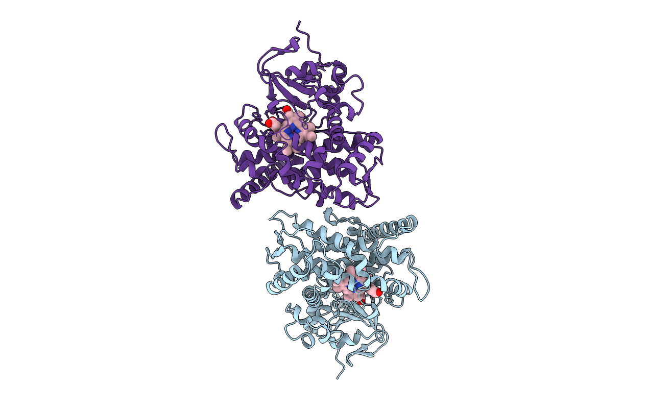
1FAH image
Entry Detail
PDB ID:
1FAH
Title:
STRUCTURE OF CYTOCHROME P450
Biological Source:
Source Organism:
Host Organism:
PDB Version:
Deposition Date:
1996-08-01
Release Date:
1997-02-12
Method Details:
Experimental Method:
Resolution:
2.30 Å
R-Value Work:
0.17
R-Value Observed:
0.17
Space Group:
P 1 21 1
Macromolecular Entities
Polymer Type:polypeptide(L)
Description:CYTOCHROME P450 BM-3
Mutations:T268A
Chain IDs:A, B
Chain Length:471
Number of Molecules:2
Biological Source:Bacillus megaterium
Ligand Molecules
Primary Citation
The role of Thr268 in oxygen activation of cytochrome P450BM-3.
Biochemistry 34 14733 14740 (1995)
PMID: 7578081 DOI: 10.1021/bi00045a014

Abstact

Cytochrome P450BM-3, a catalytically self-sufficient monooxygenase from Bacillus megaterium, catalyzes the omega-n (n = 1-3) hydroxylation of fatty acids in the presence of O2 and NADPH. Like most other P450s, cytochrome P450BM-3 contains a threonine residue (Thr268) in the distal I helix thought to be important for O2 binding and activation. Thr268 has been converted to alanine and the enzymatic properties and heme domain crystal structure determined. Using sodium laurate as the substrate, the mutant exhibited slower rates of O2 and NADPH consumption. In addition, electron transfer is uncoupled from substrate hydroxylation as evidenced by the greater production of water and peroxide in the mutant compared to the wild-type enzyme. The crystal structure of the mutant reveals that the only changes in structure are confined to the site of mutation. These data indicate an important role for Thr268 in O2 binding and activation in the metabolism of sodium laurate by cytochrome P450BM-3.

Legend

Protein

Chemical

Disease

Primary Citation of related structures