Deposition Date
2000-07-11
Release Date
2001-06-13
Last Version Date
2024-02-07
Entry Detail
PDB ID:
1F9T
Keywords:
Title:
CRYSTAL STRUCTURES OF KINESIN MUTANTS REVEAL A SIGNALLING PATHWAY FOR ACTIVATION OF THE MOTOR ATPASE
Biological Source:
Source Organism(s):
Saccharomyces cerevisiae (Taxon ID: 4932)
Expression System(s):
Method Details:
Experimental Method:
Resolution:
1.50 Å
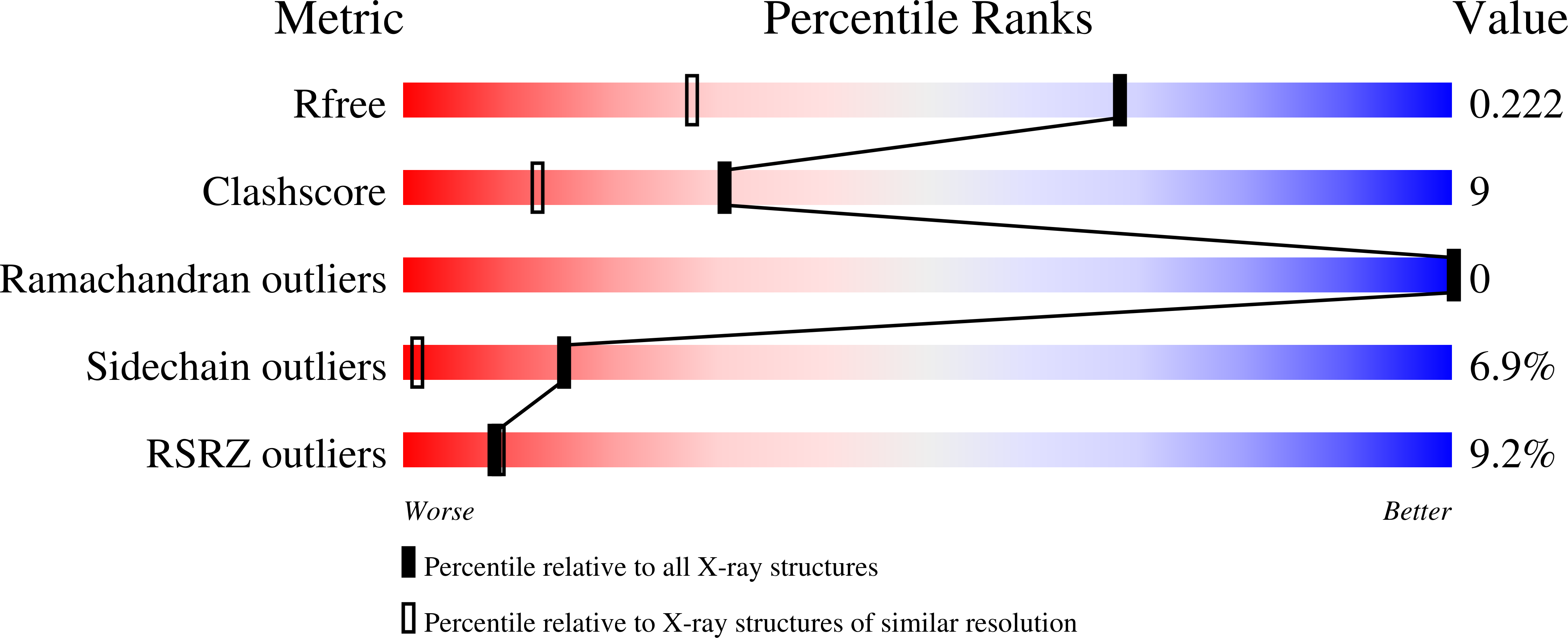
R-Value Free:
0.24
R-Value Work:
0.21
Space Group:
P 1 21 1