
Deposition Date
2000-06-30
Release Date
2001-06-30
Last Version Date
2024-10-16
Entry Detail
PDB ID:
1F8G
Keywords:
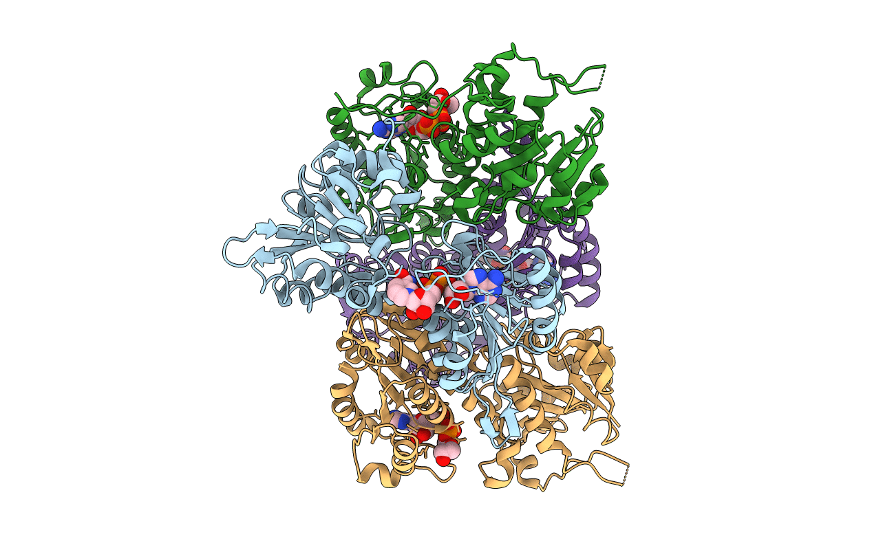
Title:
THE X-RAY STRUCTURE OF NICOTINAMIDE NUCLEOTIDE TRANSHYDROGENASE FROM RHODOSPIRILLUM RUBRUM COMPLEXED WITH NAD+
Biological Source:
Source Organism(s):
Rhodospirillum rubrum (Taxon ID: 1085)
Expression System(s):
Method Details:
Experimental Method:
Resolution:
2.00 Å
R-Value Free:
0.26
R-Value Work:
0.21
Space Group:
P 1 21 1
