1F89 image
Deposition Date 2000-06-29
Release Date 2001-10-04
Last Version Date 2024-02-07
Entry Detail
PDB ID:
1F89
Title:
Crystal structure of Saccharomyces cerevisiae Nit3, a member of branch 10 of the nitrilase superfamily
Biological Source:
Source Organism:
Host Organism:
Method Details:
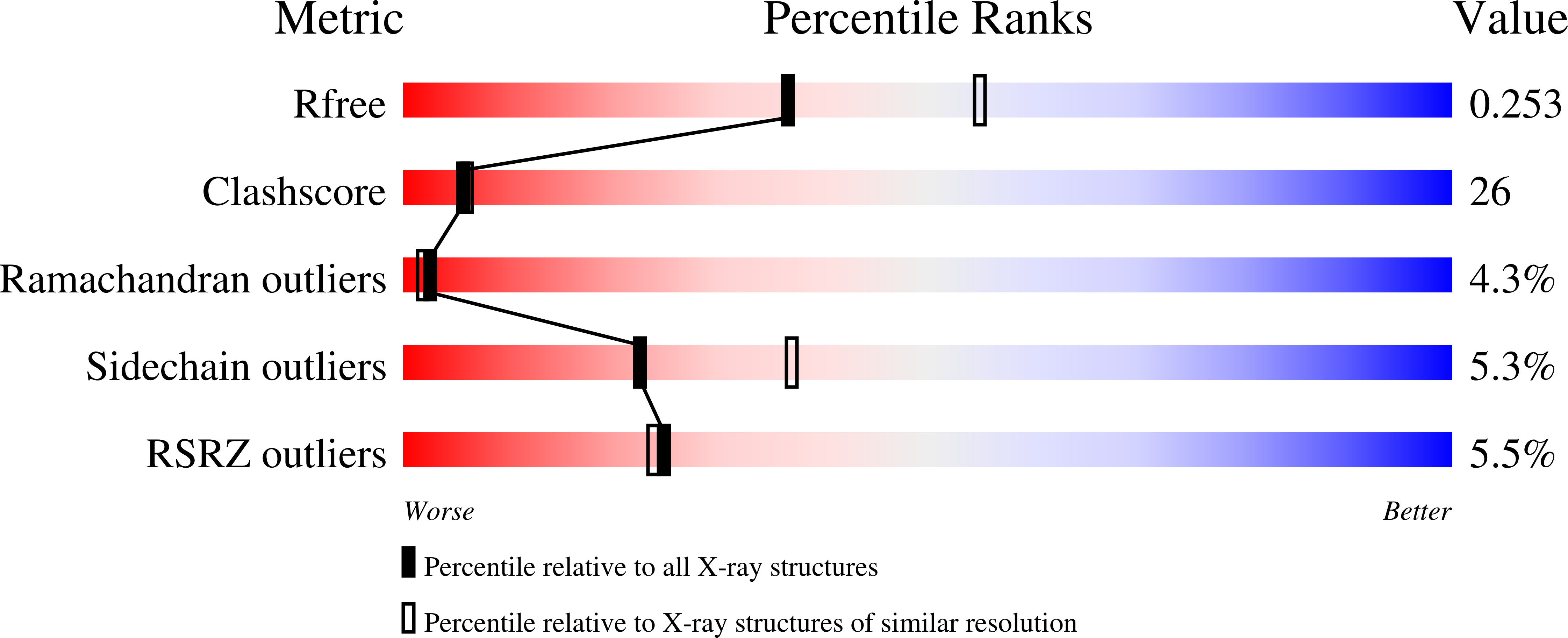
Experimental Method:
Resolution:
2.40 Å
R-Value Free:
0.25
R-Value Work:
0.22
Space Group:
P 1 21 1
Macromolecular Entities
Polymer Type:polypeptide(L)
Molecule:32.5 KDA PROTEIN YLR351C
Gene (Uniprot):NIT3
Chain IDs:A, B
Chain Length:291
Number of Molecules:2
Biological Source:Saccharomyces cerevisiae
Primary Citation
Crystal structure of a putative CN hydrolase from yeast
Proteins 52 283 291 (2003)
PMID: 12833551 DOI: 10.1002/prot.10417

Abstact

The crystal structure of a yeast hypothetical protein with sequence similarity to CN hydrolases has been determined to 2.4 A resolution by the multiwavelength anomalous dispersion (MAD) method. The protein folds as a four-layer alphabetabetaalpha sandwich and exists as a dimer in the crystal and in solution. It was selected in a structural genomics project as representative of CN hydrolases at a time when no structures had been determined for members of this family. Structures for two other members of the family have since been reported and the three proteins have similar topology and dimerization modes, which are distinct from those of other alphabetabetaalpha proteins whose structures are known. The dimers form an unusual eight-layer alphabetabetaalpha:alphabetabetaalpha structure. Although the precise enzymatic reactions catalyzed by the yeast protein are not known, considerable information about the active site may be deduced from conserved sequence motifs, comparative biochemical information, and comparison with known structures of hydrolase active sites. As with serine hydrolases, the active-site nucleophile (cysteine in this case) is positioned on a nucleophile elbow.

Legend

Protein

Chemical

Disease

Primary Citation of related structures