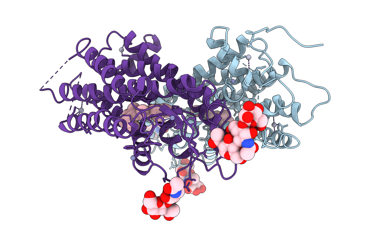
1F88 image
Deposition Date 2000-06-29
Release Date 2000-08-04
Last Version Date 2024-10-30
Entry Detail
PDB ID:
1F88
Title:
CRYSTAL STRUCTURE OF BOVINE RHODOPSIN
Biological Source:
Source Organism:
Bos taurus (Taxon ID: 9913)
Method Details:
Experimental Method:
Resolution:
2.80 Å
R-Value Free:
0.23
R-Value Work:
0.18
Space Group:
P 41
Macromolecular Entities
Polymer Type:polypeptide(L)
Molecule:RHODOPSIN
Gene (Uniprot):RHO
Chain IDs:A, B
Chain Length:348
Number of Molecules:2
Biological Source:Bos taurus
Modified Residue
Compound ID Chain ID Parent Comp ID Details 2D Image
ASN B ASN GLYCOSYLATION SITE
Primary Citation
Crystal structure of rhodopsin: A G protein-coupled receptor.
Science 289 739 745 (2000)
PMID: 10926528 DOI: 10.1126/science.289.5480.739

Abstact

Heterotrimeric guanine nucleotide-binding protein (G protein)-coupled receptors (GPCRs) respond to a variety of different external stimuli and activate G proteins. GPCRs share many structural features, including a bundle of seven transmembrane alpha helices connected by six loops of varying lengths. We determined the structure of rhodopsin from diffraction data extending to 2.8 angstroms resolution. The highly organized structure in the extracellular region, including a conserved disulfide bridge, forms a basis for the arrangement of the seven-helix transmembrane motif. The ground-state chromophore, 11-cis-retinal, holds the transmembrane region of the protein in the inactive conformation. Interactions of the chromophore with a cluster of key residues determine the wavelength of the maximum absorption. Changes in these interactions among rhodopsins facilitate color discrimination. Identification of a set of residues that mediate interactions between the transmembrane helices and the cytoplasmic surface, where G-protein activation occurs, also suggests a possible structural change upon photoactivation.

Legend

Protein

Chemical

Disease

Primary Citation of related structures