1F6M image
Deposition Date 2000-06-22
Release Date 2000-08-30
Last Version Date 2024-11-06
Entry Detail
PDB ID:
1F6M
Keywords:
Title:
CRYSTAL STRUCTURE OF A COMPLEX BETWEEN THIOREDOXIN REDUCTASE, THIOREDOXIN, AND THE NADP+ ANALOG, AADP+
Biological Source:
Source Organism:
Escherichia coli (Taxon ID: 562)
Host Organism:
Method Details:
Experimental Method:
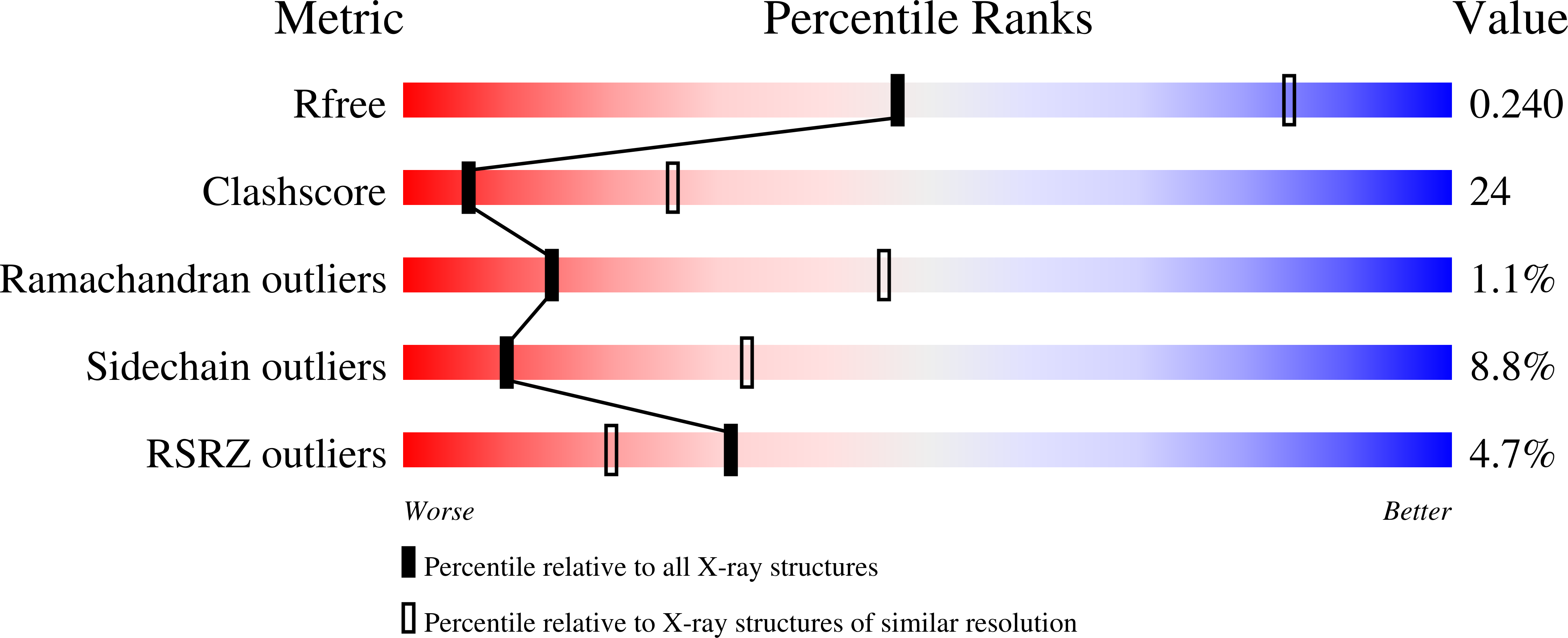
Resolution:
2.95 Å
R-Value Free:
0.24
R-Value Work:
0.20
R-Value Observed:
0.20
Space Group:
C 1 2 1
Macromolecular Entities
Polymer Type:polypeptide(L)
Molecule:THIOREDOXIN REDUCTASE
Gene (Uniprot):trxB
Mutations:C135S
Chain IDs:A, B, E, F
Chain Length:320
Number of Molecules:4
Biological Source:Escherichia coli
Polymer Type:polypeptide(L)
Molecule:THIOREDOXIN 1
Gene (Uniprot):trxA
Mutations:C35S
Chain IDs:C, D, G, H
Chain Length:108
Number of Molecules:4
Biological Source:Escherichia coli
Primary Citation
Twists in catalysis: alternating conformations of Escherichia coli thioredoxin reductase.
Science 289 1190 1194 (2000)
PMID: 10947986 DOI: 10.1126/science.289.5482.1190

Abstact

In thioredoxin reductase (TrxR) from Escherichia coli, cycles of reduction and reoxidation of the flavin adenine dinucleotide (FAD) cofactor depend on rate-limiting rearrangements of the FAD and NADPH (reduced form of nicotinamide adenine dinucleotide phosphate) domains. We describe the structure of the flavin-reducing conformation of E. coli TrxR at a resolution of 3.0 angstroms. The orientation of the two domains permits reduction of FAD by NADPH and oxidation of the enzyme dithiol by the protein substrate, thioredoxin. The alternate conformation, described by Kuriyan and co-workers, permits internal transfer of reducing equivalents from reduced FAD to the active-site disulfide. Comparison of these structures demonstrates that switching between the two conformations involves a "ball-and-socket" motion in which the pyridine nucleotide-binding domain rotates by 67 degrees.

Legend

Protein

Chemical

Disease

Primary Citation of related structures